File:Keibel Mall 2 022.jpg
Original file (821 × 950 pixels, file size: 113 KB, MIME type: image/jpeg)
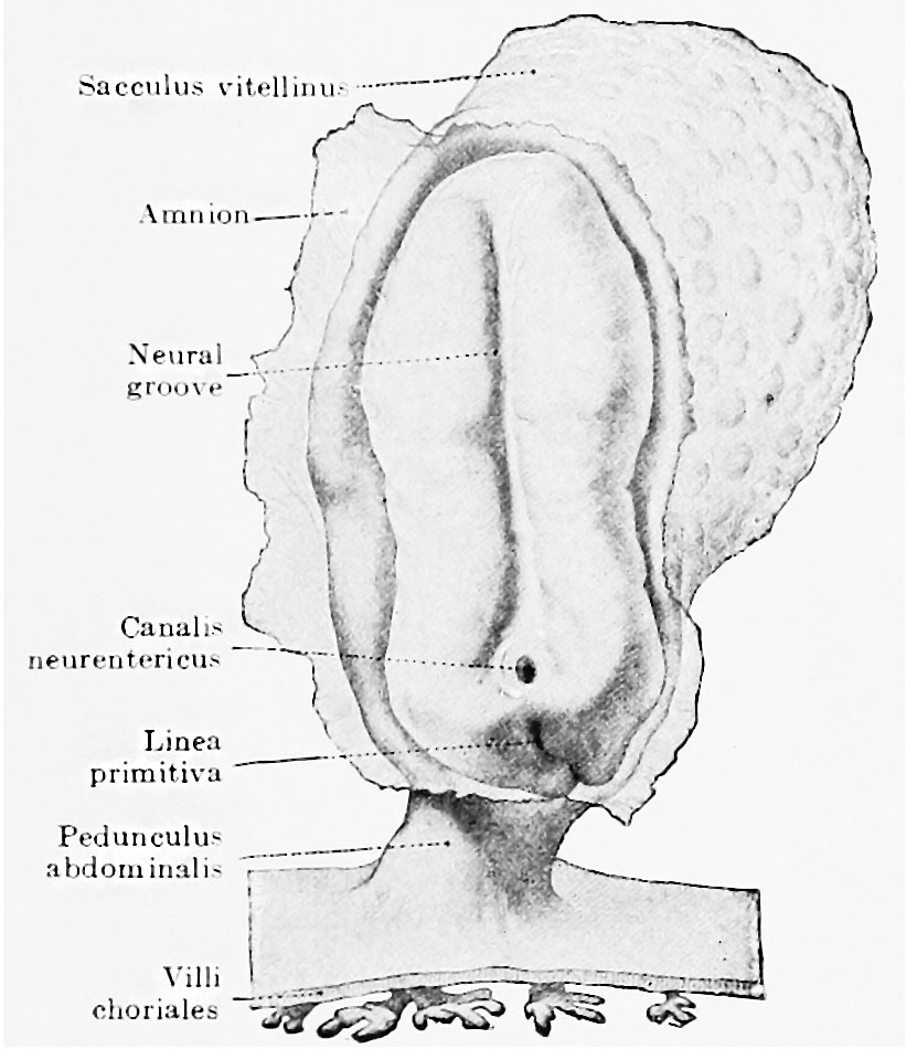
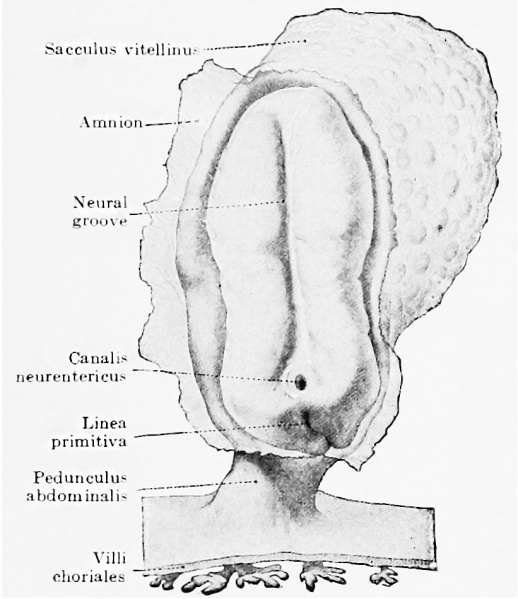
Fig. 22. Dorsal view of human embryo in the neural-plate stage
The amnion is opened, exposing the germinal plate lying directly on the yolk-sac. (Graf Spee, from Kollmann.) The further elevation of the edges and their approximation and fusion across the median line to form the neural tube is shown in four stages in Fig. 23. The comparison of these figures shows that the formation of the neural tube is most advanced in the middle of the germ plate corresponding to the junction of brain and spinal cord. From this region the differentiation and closure of the tube extends caudally and orally, the last portions to close being called the anterior and posterior neuropores (see Figs. 24 and 26). The process of closure, though it always begins in about the same region, shows some variation in the time of its occurrence. In Fig. 23, C, it is further advanced than in Fig. 23, D, which, judging from the number of somites, is the older embryo of the two.
| Historic Disclaimer - information about historic embryology pages |
|---|
 |
CNS Figures: 22 - neural-plate stage | 23 - 5 to 14 somites stage | 24 - 3.2 mm embryo | 30 - brain 3, 4 and 8 weeks | 32 - 10 mm cranial nerves
- XIV. The Development of the Nervous System: The Histogenesis of Nervous Tissue | The Development of the Central Nervous System | The Peripheral Nervous System | The Sympathetic Nervous System | Manual of Human Embryology II
Cite this page: Hill, M.A. (2024, April 16) Embryology Keibel Mall 2 022.jpg. Retrieved from https://embryology.med.unsw.edu.au/embryology/index.php/File:Keibel_Mall_2_022.jpg
- © Dr Mark Hill 2024, UNSW Embryology ISBN: 978 0 7334 2609 4 - UNSW CRICOS Provider Code No. 00098G
File history
Click on a date/time to view the file as it appeared at that time.
| Date/Time | Thumbnail | Dimensions | User | Comment | |
|---|---|---|---|---|---|
| current | 10:48, 8 October 2018 | | 821 × 950 (113 KB) | Z8600021 (talk | contribs) | |
| 10:47, 8 October 2018 | 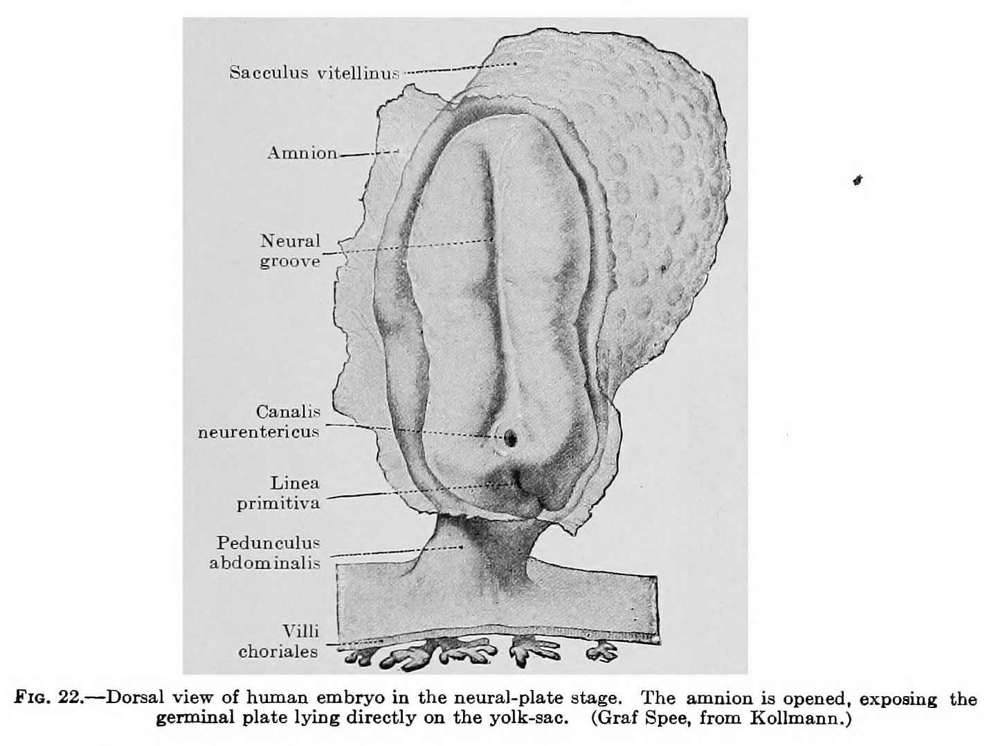 | 1,439 × 1,078 (190 KB) | Z8600021 (talk | contribs) |
You cannot overwrite this file.
File usage
The following 2 pages use this file: