File:Keibel Mall 2 001.jpg

Original file (800 × 733 pixels, file size: 135 KB, MIME type: image/jpeg)
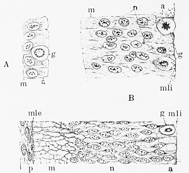
Fig. 1. Three early stages in the development of the wall of the neural tube
Showing the conversion of the single layer of discrete cells into a richly nucleated syncytial framework.
A, medullary plate of embryo rabbit just before closure of neural tube;
B, similar section of 5 mm. pig embryo just after closure of neural tube;
C, wall of neural tube of 10 mm. pig embryo;
a, ependymal layer; g, germinal cell; m, marginal layer; mle and mli, external and internal limiting membranes; n, mantle or nuclear layer; p, mesoderm.
(After Hardesty.)
Hardesty I. On the development and nature of the neuroglia. (1904) Amer. J Anat. 3.
Hardesty I. On the occurrence of sheath cells and the nature of the axone sheaths in the central nervous system. (1904) Amer. J Anat. 4.
File history
Click on a date/time to view the file as it appeared at that time.
| Date/Time | Thumbnail | Dimensions | User | Comment | |
|---|---|---|---|---|---|
| current | 09:06, 3 March 2017 | | 800 × 733 (135 KB) | Z8600021 (talk | contribs) | |
| 09:06, 3 March 2017 |  | 1,414 × 1,251 (324 KB) | Z8600021 (talk | contribs) | Fig. 1. Three early stages in the development of the wall of the neural tube, |
You cannot overwrite this file.
File usage
The following 2 pages use this file: