File:Keibel Mall 248-259.jpg

Original file (709 × 1,108 pixels, file size: 189 KB, MIME type: image/jpeg)
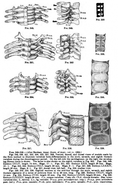
Figs. 248-259.— (After flardeen. Anier. Journ. of Anal,, vol. iv. 1805.) Figs. 248. 249, 251, 252, 254. 255, 257, 258. Ventral, lateral, and doraal views of model made by vertebrae during the chandrogenoui" period. On the left side the cartllaginouii, on the right the enveloping fibrous liwue i* shown. The latter is also shown on the eighth vertebra in Fig. 252. Fign. 248. 249. Embrj'o CXI.IV, lengtli 14 mm. M«gn. 17 : 1. Figs. 251. 252. Embryo XXII. length 20 mm. Magn. 13:1. Figs. 254. 255. Embryo CXLV. length 33 mm. Magn.QM. Figs. 2S7, 258. Embryo LXXXIV, length 50 mm. Magn. 9:1. Fig. 257. dorsal view, left half . Fig. 258. median view. Figs. 250, 253, 250. 259. sagittal -enions in the mid-line through the niilh, seventh, and eighth thoracic segments of a series of embryos from 15 to 50 mm. long. Fig. 250. Embryo CXLI\'. length 14 mm. Fig. 25B. Embryo C\'1I1, length 22 mm. Fig. 256. Embryo LXXIX. lenglh 33 mm. Fig. 259. Embryo CLXXXlV. length 50mm, C.e.. eorpus vertebra; Co«o, rib; CA.rf., chorda dorsalis; Z>t«, inteiw vertebtaldise; I,., lamina; L.v.. lig. ventrale; /'r.a.a.. proc.articulariaant. Isup,); Prji.p., proe. articularia post. {iaf.J; Pr.n.. proc. neumlis; Fr.rd., prae. radicularit; Pr.t.. prnc. spinaliK; PrJr., proc. transversus.
- KM Figure Links: The Germ Cells | Segmentation | First Primitive Segment | Gastrulation | External Form | Placenta | Axial Skeleton | Limb Skeleton | Skull | Muscular System
| Historic Disclaimer - information about historic embryology pages |
|---|
 |
Glossary Links
- Glossary: A | B | C | D | E | F | G | H | I | J | K | L | M | N | O | P | Q | R | S | T | U | V | W | X | Y | Z | Numbers | Symbols | Term Link
Cite this page: Hill, M.A. (2024, April 25) Embryology Keibel Mall 248-259.jpg. Retrieved from https://embryology.med.unsw.edu.au/embryology/index.php/File:Keibel_Mall_248-259.jpg
- © Dr Mark Hill 2024, UNSW Embryology ISBN: 978 0 7334 2609 4 - UNSW CRICOS Provider Code No. 00098G
File history
Click on a date/time to view the file as it appeared at that time.
| Date/Time | Thumbnail | Dimensions | User | Comment | |
|---|---|---|---|---|---|
| current | 22:48, 2 October 2012 | | 709 × 1,108 (189 KB) | Z8600021 (talk | contribs) |
You cannot overwrite this file.
File usage
The following 2 pages use this file: