File:Keibel Mall 038-039.jpg
Keibel_Mall_038-039.jpg (600 × 389 pixels, file size: 30 KB, MIME type: image/jpeg)
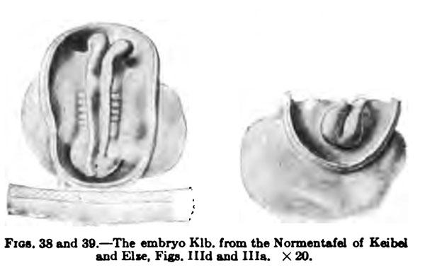
Fig. 38. and 39.
I place next the embryo Klb. of the Normentafel of Keibel and Elze, Figs. Hid and Ilia.
The ovum which contained the embryo was obtained by a laparotomy and both it and the embryo may be regarded as quite normal. The embryo had five to six pairs of primitive somites. The vertex bend had already begun to form, otherwise the embryo lies flat on the yolk sack; it has no dorsal bend. Fig. 38 shows it from the dorsal surface, and, the amnion being cut away, it is seen to be attached to the chorion, a small portion of which is represented, by a short belly stalk. To the right and left of the cut edges of the amnion the yolk sack projects beyond the embryonic structures. Both the cranial and caudal ends of the embryo are separated from the yolk sack, but the middle portion is still spread out. The medullary groove is widely open, but rather deep. Caudally the medullary folds embrace the dorsal opening of the neurenteric canal and the cranial end of the primitive streak, which immediately bends downward so that it cannot be perceived in its entire extent; indeed, it lias already undergone much retrogression. The well-marked medullary anlage shows the vertex bend, so that its cranial end cannot be seen in a dorsal view. The brain part of the medullary anlage shows a separation into three portions, the most caudal of which extends to about the fourth pair of primitive somites and then passes without any marked boundary into the spinal cord. I may here note that by far the greater part of the dorsal region of this embryo belongs to the later head region. The boundary between the head and trunk regions passes through the fourth primitive somite. The most anterior somite of the neck region is first differentiated.
Fig. 39 shows the embryo from in front. The amnion has again been removed; ventrally the relatively large yolk sack is seen. Of the embrjonic anlage only the ventrally bent portion, as far back as about the vertex bend, can be seen; but it may be observed that the most anterior portion of the brain anlage is relatively greatly developed. The medullary groove terminates a little behind the cranial end of the medullary aniage, so that the latter shows anteriorly a transverse ridge. Nothing is yet to be seen of the optic pits, the forerunners of the optic vesicles.
File history
Click on a date/time to view the file as it appeared at that time.
| Date/Time | Thumbnail | Dimensions | User | Comment | |
|---|---|---|---|---|---|
| current | 15:13, 15 February 2012 |  | 600 × 389 (30 KB) | S8600021 (talk | contribs) |
You cannot overwrite this file.
File usage
The following 2 pages use this file:
