File:Keibel1910 fig24.jpg
Keibel1910_fig24.jpg (520 × 520 pixels, file size: 25 KB, MIME type: image/jpeg)
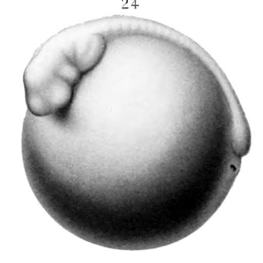
Fig. 24. Dorso-lateral view of egg 22 days 17 hrs old
Dorso-lateral view of embryo 22 days 17 hrs. old, length 8 mm, l6~i8 pairs of myotomes. Embryo much curved laterally. Anterior half of head free from yolk. Caudal enlargement more prominent. Optic vesicles and mandibular arch well defined. The hyoid and first branchial arches are discernible ; also the common anläge of the second and third branchial arches. (X 10.)
| Historic Disclaimer - information about historic embryology pages |
|---|
| Pages where the terms "Historic" (textbooks, papers, people, recommendations) appear on this site, and sections within pages where this disclaimer appears, indicate that the content and scientific understanding are specific to the time of publication. This means that while some scientific descriptions are still accurate, the terminology and interpretation of the developmental mechanisms reflect the understanding at the time of original publication and those of the preceding periods, these terms, interpretations and recommendations may not reflect our current scientific understanding. (More? Embryology History | Historic Embryology Papers) |
Reference
Eycleshymer AC. and Wilson JM. Normal Plates of the Development of the Salamander Embryo (Nectürüs maculosus). Vol. 11 in series by Keibel F. Normal plates of the development of vertebrates (Normentafeln zur Entwicklungsgeschichte der Wirbelthiere) Fisher, Jena., Germany.
Cite this page: Hill, M.A. (2024, April 20) Embryology Keibel1910 fig24.jpg. Retrieved from https://embryology.med.unsw.edu.au/embryology/index.php/File:Keibel1910_fig24.jpg
- © Dr Mark Hill 2024, UNSW Embryology ISBN: 978 0 7334 2609 4 - UNSW CRICOS Provider Code No. 00098G
File history
Click on a date/time to view the file as it appeared at that time.
| Date/Time | Thumbnail | Dimensions | User | Comment | |
|---|---|---|---|---|---|
| current | 14:11, 10 January 2015 |  | 520 × 520 (25 KB) | Z8600021 (talk | contribs) | ==Fig. 24. Dorso-lateral view of egg 22 days 17 hrs old== Dorso-lateral view of embryo 22 days 17 hrs. old, length 8 mm, l6~i8 pairs of myotomes. Embryo much curved laterally. Anterior half of head free from yolk. Caudal enlargement more prominent. Op... |
You cannot overwrite this file.
File usage
The following 3 pages use this file:

