File:Johnston1907 fig019.jpg
From Embryology
Size of this preview: 541 × 600 pixels. Other resolution: 1,280 × 1,419 pixels.
Original file (1,280 × 1,419 pixels, file size: 236 KB, MIME type: image/jpeg)
Summary
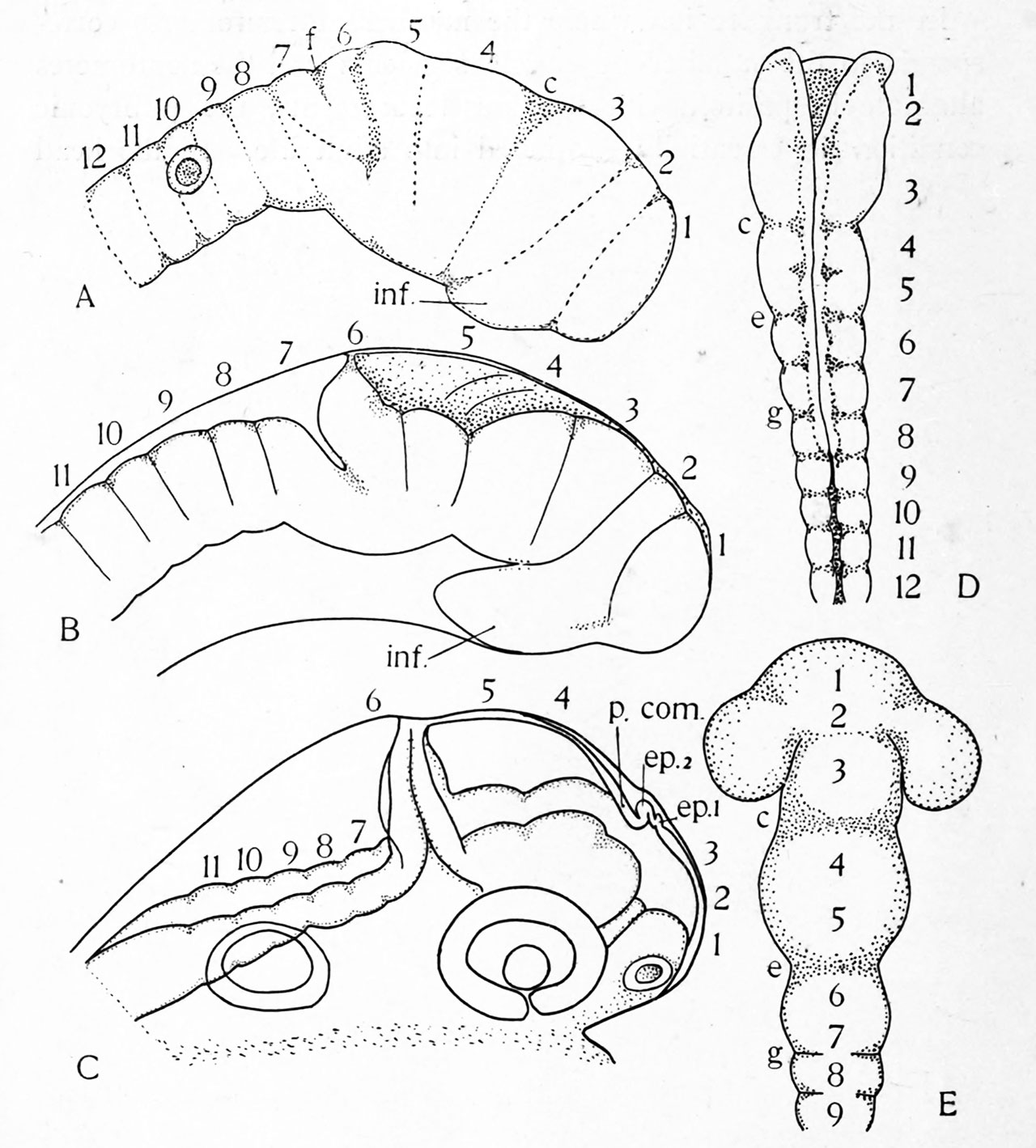
Fig. 19. The history of the neuromeres in the bony fish and the chick. A, B, C, three stages in the development of the bony fish, as seen from the right side. D, E, the brain of the chick. After Hill. The letters c. e. g. mark corresponding furrows in the chick brain; ep. i, ep. 2, anterior and posterior epiphyses; in}, inferior lobe; p. com., posterior commissure. The neuromeres are indicated by Arabic numerals.
File history
Click on a date/time to view the file as it appeared at that time.
| Date/Time | Thumbnail | Dimensions | User | Comment | |
|---|---|---|---|---|---|
| current | 22:48, 23 February 2020 | | 1,280 × 1,419 (236 KB) | Z8600021 (talk | contribs) | |
| 22:46, 23 February 2020 |  | 2,169 × 2,762 (647 KB) | Z8600021 (talk | contribs) | Fig. 19. The history of the neuromeres in the bony fish and the chick. A, B, C, three stages in the development of the bony fish, as seen from the right side. D, E, the brain of the chick. After Hill. The letters c. e. g. mark corresponding furrows in the chick brain; ep. i, ep. 2, anterior and posterior epiphyses; in}, inferior lobe; p. com., posterior commissure. The neuromeres are indicated by Arabic numerals. |
You cannot overwrite this file.
File usage
The following 2 pages use this file: