File:Jackson1909a fig01.jpg
Original file (1,280 × 1,318 pixels, file size: 192 KB, MIME type: image/jpeg)
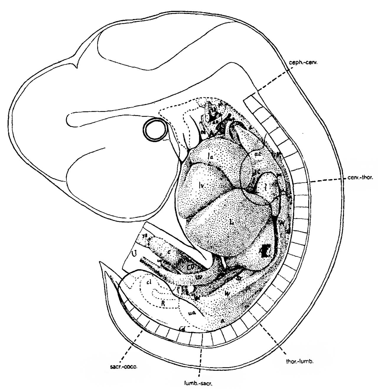
Fig. 1. Graphic recontruction of an 11 mm Human Embryo
(No. 60) from the left side, showing the body outline. extremities, central nervous system, vertebral centra1, viscera, etc. The parts corresponding to the viscera in the model (Fig. 5) are indicated by stippling. The various regions of the vertebral column are indicated (ceph.-cerv., cerv.-thor., thor.-lumb.. lumb. sacr., sacr.-cocc.).
A, ascending aorta; a, descending aorta; a3, a4, 3d and 4th aortic arches: ac. anterior cardinal (jugular) vein; c, anlage of the cocum; cd, caudal aorta; cl, cloaca; co. colon; dC, ductus Cuvieri; l, lung; L, liver; in, left auricle; lv, left ventricle; pc, posterior cardinal vein; ph. pharynx; R, rectum; sx, sexual anlage; sl, left suprarenal anlnge; sa, origin of subclavian artery: th, anluge of thymus; tl. tm, lateral and median anlagms of the thyroid: U, umbilical cord: ua, umbilical artery; uv. umbilical vein; w. Wolffian Body; wd. Wolffian duct; x, wlndow cut into great omeutum: ys, attachment of yolk-stalk to intestinal loop.
| Historic Disclaimer - information about historic embryology pages |
|---|
 |
- Jackson 1909 Figures: Fig 1. 11 mm embryo | Fig 2. 17 mm embryo | Fig 3. 31 mm embryo | Fig 4. 65 mm embryo | Fig. 5-8 | Fig 5. 11 mm embryo | Fig 6. 17 mm embryo | Fig 7. 31 mm embryo | Fig 8. 65 mm embryo

Fig 1. 11 mm embryo
Fig 2. 17 mm embryo
Fig 3. 31 mm embryo
Fig 4. 65 mm embryo
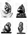
Fig. 5-8.
Fig 5. 11 mm embryo
Fig 6. 17 mm embryo
Fig 7. 31 mm embryo
Fig 8. 65 mm embryo


Reference
Jackson CM. On the developmental topography of the thoracic and abdominal viscera. (1909) Anat. Rec. 111: -396.
Cite this page: Hill, M.A. (2024, April 23) Embryology Jackson1909a fig01.jpg. Retrieved from https://embryology.med.unsw.edu.au/embryology/index.php/File:Jackson1909a_fig01.jpg
- © Dr Mark Hill 2024, UNSW Embryology ISBN: 978 0 7334 2609 4 - UNSW CRICOS Provider Code No. 00098G
File history
Click on a date/time to view the file as it appeared at that time.
| Date/Time | Thumbnail | Dimensions | User | Comment | |
|---|---|---|---|---|---|
| current | 10:33, 22 February 2018 | | 1,280 × 1,318 (192 KB) | Z8600021 (talk | contribs) | |
| 10:30, 22 February 2018 | 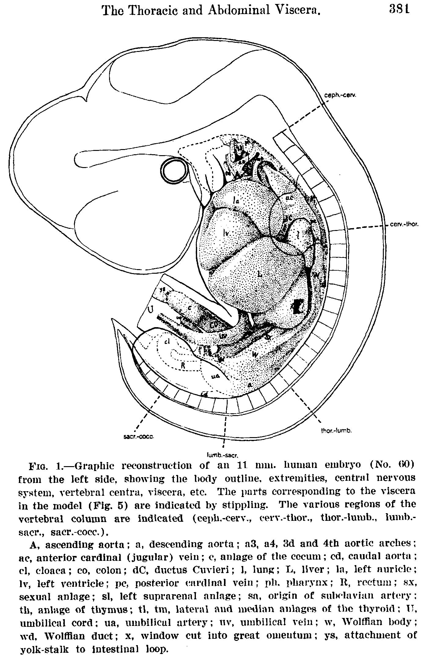 | 1,417 × 2,177 (446 KB) | Z8600021 (talk | contribs) | {{Jackson1909a figures}} |
You cannot overwrite this file.