File:Ingalls1932b fig02.jpg
Original file (415 × 653 pixels, file size: 59 KB, MIME type: image/jpeg)
Summary
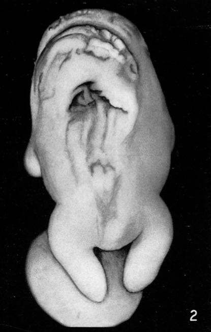
Fig. 2. Embryo No. 46
Greatest length 14.5mm. The entire body is very much malformed. Almost the whole of the dorsum is markedly altered or defective. The deep triangular cavity is due to the postmortem loss of tissue.
Reference
Ingalls NW. Studies in the pathology of development: II. Some aspects of defective development in the dorsal midline. (1932) Am J Pathol. 8(5): 525-556 PMID 19970035
Cite this page: Hill, M.A. (2024, April 19) Embryology Ingalls1932b fig02.jpg. Retrieved from https://embryology.med.unsw.edu.au/embryology/index.php/File:Ingalls1932b_fig02.jpg
- © Dr Mark Hill 2024, UNSW Embryology ISBN: 978 0 7334 2609 4 - UNSW CRICOS Provider Code No. 00098G
File history
Click on a date/time to view the file as it appeared at that time.
| Date/Time | Thumbnail | Dimensions | User | Comment | |
|---|---|---|---|---|---|
| current | 09:14, 14 October 2020 | | 415 × 653 (59 KB) | Z8600021 (talk | contribs) | ==Fig. 2. Embryo No. 46== Greatest length 14.5mm. The entire body is very much malformed. Almost the whole of the dorsum is markedly altered or defective. The deep triangular cavity is due to the postmortem loss of tissue. ===Reference=== {{Ref-Ingalls1932b}} {{footer}} |
You cannot overwrite this file.
File usage
The following 2 pages use this file: