File:Ingalls1918 plate 3.jpg

Original file (905 × 1,200 pixels, file size: 132 KB, MIME type: image/jpeg)
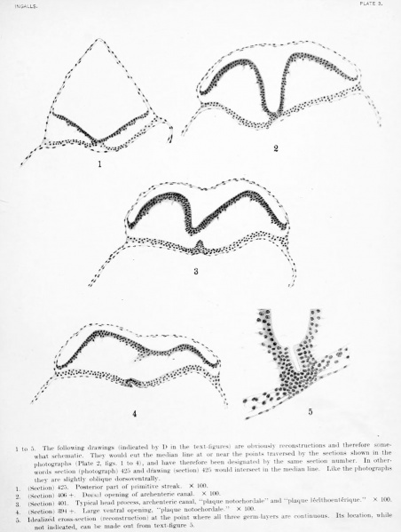
Plate 3
1 to 5. The following drawings (indicated by D in the text-figures) are obviously reconstructions and therefore somewhat schematic. They would cut the median line at or near the points traversed by the sections shown in the photographs (Plate 2. figs. 1 to 4), and have therefore been designated by the same section number. In otherwords section (photograph) 425 and drawing (section) 425 would intereect in tlie median line. Like the photographs ihey are slightly oblique dorsoventrally.
- (Section) 425. Posterior part of primitive streak. X 100.
- (Section) 406 +. Dorsal opening of archenteric canal. X 100.
- (Section) 401. Typical head process, archenteric canal, "plaque notochordale' and "plaque lec'ithoenterique." X 100.
- (Section) 394 +. Large ventral opening, "plaque notochordale." X 100.
- Idealized cross-section (reconstruction) at the point where all three germ-layers; are continuous. Its location, while not indicated, can be made out from text-figure 5.
- Contribution No.23: Figures | Plate 1 | Plate 2 | Plate 3 | Plate 4 | Plate 1 | Carnegie - Contributions to Embryology | Carnegie stage 8 | Category:Carnegie Stage 8 | Historic Embryology Papers
| Historic Disclaimer - information about historic embryology pages |
|---|
| Pages where the terms "Historic" (textbooks, papers, people, recommendations) appear on this site, and sections within pages where this disclaimer appears, indicate that the content and scientific understanding are specific to the time of publication. This means that while some scientific descriptions are still accurate, the terminology and interpretation of the developmental mechanisms reflect the understanding at the time of original publication and those of the preceding periods, these terms, interpretations and recommendations may not reflect our current scientific understanding. (More? Embryology History | Historic Embryology Papers) |
Reference
Ingalls NW. A human embryo before the appearance of the myotomes. (1918) Contrib. Embryol., Carnegie Inst. Wash. No.23 Publ. 227, 7:111-134.
Cite this page: Hill, M.A. (2024, April 20) Embryology Ingalls1918 plate 3.jpg. Retrieved from https://embryology.med.unsw.edu.au/embryology/index.php/File:Ingalls1918_plate_3.jpg
- © Dr Mark Hill 2024, UNSW Embryology ISBN: 978 0 7334 2609 4 - UNSW CRICOS Provider Code No. 00098G
| Historic Disclaimer - information about historic embryology pages |
|---|
| Pages where the terms "Historic" (textbooks, papers, people, recommendations) appear on this site, and sections within pages where this disclaimer appears, indicate that the content and scientific understanding are specific to the time of publication. This means that while some scientific descriptions are still accurate, the terminology and interpretation of the developmental mechanisms reflect the understanding at the time of original publication and those of the preceding periods, these terms, interpretations and recommendations may not reflect our current scientific understanding. (More? Embryology History | Historic Embryology Papers) |
File history
Click on a date/time to view the file as it appeared at that time.
| Date/Time | Thumbnail | Dimensions | User | Comment | |
|---|---|---|---|---|---|
| current | 01:51, 28 December 2012 |  | 905 × 1,200 (132 KB) | Z8600021 (talk | contribs) |
You cannot overwrite this file.
File usage
The following page uses this file:
