File:Ingalls1918 plate 1 fig4+5.jpg
From Embryology
Size of this preview: 800 × 538 pixels. Other resolution: 1,000 × 672 pixels.
Original file (1,000 × 672 pixels, file size: 161 KB, MIME type: image/jpeg)
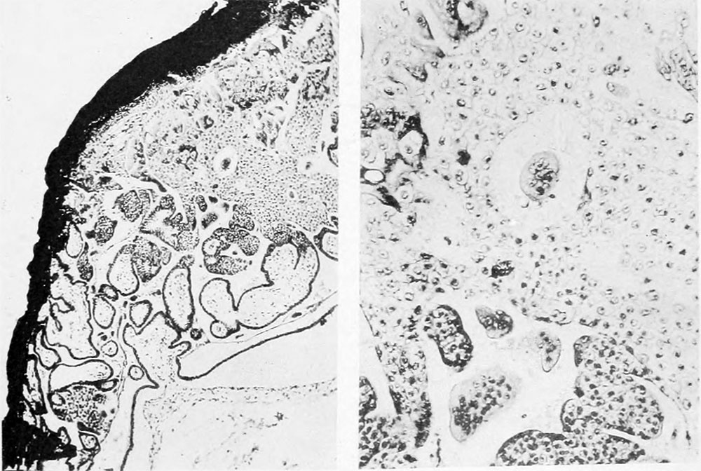
Plate 1
1 and 2. Intact vesicle, opposite views. X 4.5
3. Photograph of Section 404. X 9
4. Detail of Fig. 3 (lower left corner). X 45).
5. Detail of Fig. 4. X I80.
- Contribution No.23: Figures | Plate 1 | Plate 2 | Plate 3 | Plate 4 | Plate 1 | Carnegie - Contributions to Embryology | Carnegie stage 8 | Category:Carnegie Stage 8 | Historic Embryology Papers
| Historic Disclaimer - information about historic embryology pages |
|---|
 |
Reference
Ingalls NW. A human embryo before the appearance of the myotomes. (1918) Contrib. Embryol., Carnegie Inst. Wash. No.23 Publ. 227, 7:111-134.
Cite this page: Hill, M.A. (2024, April 18) Embryology Ingalls1918 plate 1 fig4+5.jpg. Retrieved from https://embryology.med.unsw.edu.au/embryology/index.php/File:Ingalls1918_plate_1_fig4%2B5.jpg
- © Dr Mark Hill 2024, UNSW Embryology ISBN: 978 0 7334 2609 4 - UNSW CRICOS Provider Code No. 00098G
File history
Click on a date/time to view the file as it appeared at that time.
| Date/Time | Thumbnail | Dimensions | User | Comment | |
|---|---|---|---|---|---|
| current | 11:29, 2 January 2013 | | 1,000 × 672 (161 KB) | Z8600021 (talk | contribs) | ==Plate 1== 1 and 2. Intact vesicle, opposite views. X 4.5 3. Photograph of Section 404. X 9 4. Detail of Fig. 3 (lower left corner). X 45). 5. Detail of Fig. 4. X I80. {{Ingalls1918 links}} {{Historic Disclaimer}} |
You cannot overwrite this file.
File usage
The following page uses this file: