File:Ingalls1918 fig04.jpg
Original file (1,027 × 800 pixels, file size: 73 KB, MIME type: image/jpeg)
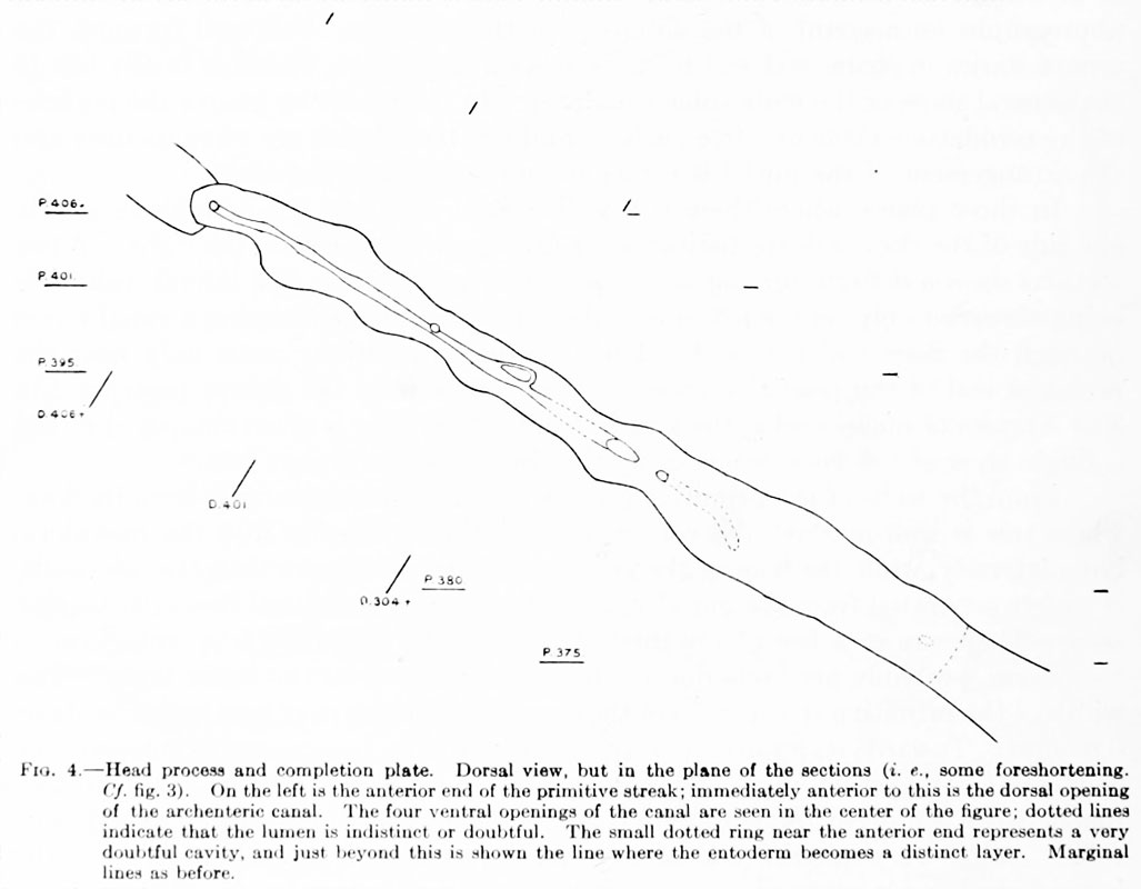
Fig. 4. Head process and completion plate
Dorsal view, but in the plane of the sections (i.e., some foreshortening. Cf. fig. 3). On the left is the anterior end of the primitive streak; immediately anterior to this is the dorsal opening of the archenteric canal. The four ventral openings of the canal arc seen in the center of the figure: dotted lines indicate that the lumen is indistinct or doubtful. The small dotted ring near the anterior end represents a very doubtful cavity, and just beyond this is shown the line where the entoderm becomes a distinct layer. Marginal lines as before.
File history
Click on a date/time to view the file as it appeared at that time.
| Date/Time | Thumbnail | Dimensions | User | Comment | |
|---|---|---|---|---|---|
| current | 01:29, 28 December 2012 | | 1,027 × 800 (73 KB) | Z8600021 (talk | contribs) | ==Fig. 4 Head process and completion plate== Right lateral view. The primitive streak, on the left, and the posterior end of the archenteric canal are represented as being in line with the more anterior structures. Other explanations under fig. 4. |
You cannot overwrite this file.
File usage
The following page uses this file: