File:In vitro fetal palate explant culture.jpg
In_vitro_fetal_palate_explant_culture.jpg (506 × 519 pixels, file size: 213 KB, MIME type: image/jpeg)
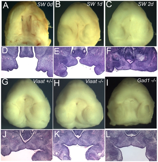
(A-F) Wild-type palates from Swiss Webster (SW) embryos at E13.5 were dissected and cultured for 2 days. (A-C) View of the oral surface of explants prior to culture (A) and after 1 day (B) or 2 days (C) of culture. (D-F) sections through the palate explants in shown in panels A-C. (G-I) Normal palatogenesis of the Viaat heterozygous (+/−) (G) and homozygous (−/−) (H) explants after 2 days in culture. Palate explants from the Gad1 mutant embryo at E13.5 also developed normally during culture for 2 days (I). (J-L) Sections of the palate explants shown in panels G-I.[1] [1]
Reference
- ↑ <pubmed>PMC2841638</pubmed>
Copyright Oh et al. This is an open-access article distributed under the terms of the Creative Commons Attribution License, which permits unrestricted use, distribution, and reproduction in any medium, provided the original author and source are credited.
File history
Click on a date/time to view the file as it appeared at that time.
| Date/Time | Thumbnail | Dimensions | User | Comment | |
|---|---|---|---|---|---|
| current | 03:21, 18 August 2011 | | 506 × 519 (213 KB) | Z3272325 (talk | contribs) | (A-F) Wild-type palates from Swiss Webster (SW) embryos at E13.5 were dissected and cultured for 2 days. (A-C) View of the oral surface of explants prior to culture (A) and after 1 day (B) or 2 days (C) of culture. (D-F) sections through the palate explan |
You cannot overwrite this file.
File usage
The following 2 pages use this file: