File:Image1184.jpg
Image1184.jpg (800 × 378 pixels, file size: 36 KB, MIME type: image/jpeg)
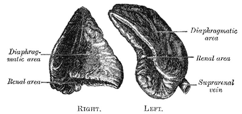
Adrenal (Suprarenal) glands viewed from behind
Relations.—The relations of the suprarenal glands differ on the two sides of the body.
The right suprarenal is situated behind the inferior vena cava and right lobe of the liver, and in front of the diaphragm and upper end of the right kidney. It is roughly triangular in shape; its base, directed downward, is in contact with the medial and anterior aspects of the upper end of the right kidney. It presents two surfaces for examination, an anterior and a posterior. The anterior surface looks forward and lateralward, and has two areas: a medial, narrow, and non-peritoneal, which lies behind the inferior vena cava; and a lateral, somewhat triangular, in contact with the liver. The upper part of the latter surface is devoid of peritoneum, and is in relation with the bare area of the liver near its lower and medial angle, while its inferior portion is covered by peritoneum, reflected onto it from the inferior layer of the coronary ligament; occasionally the duodenum overlaps the inferior portion. A little below the apex, and near the anterior border of the gland, is a short furrow termed the hilum, from which the suprarenal vein emerges to join the inferior vena cava. The posterior surface is divided into upper and lower parts by a curved ridge: the upper, slightly convex, rests upon the diaphragm; the lower, concave, is in contact with the upper end and the adjacent part of the anterior surface of the kidney.
The left suprarenal, slightly larger than the right, is crescentic in shape, its concavity being adapted to the medial border of the upper part of the left kidney. It presents a medial border, which is convex, and a lateral, which is concave; its upper end is narrow, and its lower rounded. Its anterior surface has two areas: an upper one, covered by the peritoneum of the omental bursa, which separates it from the cardiac end of the stomach, and sometimes from the superior extremity of the spleen; and a lower one, which is in contact with the pancreas and lienal artery, and is therefore not covered by the peritoneum. On the anterior surface, near its lower end, is a furrow or hilum, directed downward and forward, from which the suprarenal vein emerges. Its posterior surface presents a vertical ridge, which divides it into two areas; the lateral area rests on the kidney, the medial and smaller on the left crus of the diaphragm.
- Links: Adrenal Development
- Gray's Images: Development | Lymphatic | Neural | Vision | Hearing | Somatosensory | Integumentary | Respiratory | Gastrointestinal | Urogenital | Endocrine | Surface Anatomy | iBook | Historic Disclaimer
| Historic Disclaimer - information about historic embryology pages |
|---|
| Pages where the terms "Historic" (textbooks, papers, people, recommendations) appear on this site, and sections within pages where this disclaimer appears, indicate that the content and scientific understanding are specific to the time of publication. This means that while some scientific descriptions are still accurate, the terminology and interpretation of the developmental mechanisms reflect the understanding at the time of original publication and those of the preceding periods, these terms, interpretations and recommendations may not reflect our current scientific understanding. (More? Embryology History | Historic Embryology Papers) |
| iBook - Gray's Embryology | |
|---|---|

|
|
Reference
Gray H. Anatomy of the human body. (1918) Philadelphia: Lea & Febiger.
Cite this page: Hill, M.A. (2024, April 23) Embryology Image1184.jpg. Retrieved from https://embryology.med.unsw.edu.au/embryology/index.php/File:Image1184.jpg
- © Dr Mark Hill 2024, UNSW Embryology ISBN: 978 0 7334 2609 4 - UNSW CRICOS Provider Code No. 00098G
File history
Click on a date/time to view the file as it appeared at that time.
| Date/Time | Thumbnail | Dimensions | User | Comment | |
|---|---|---|---|---|---|
| current | 17:25, 16 May 2012 |  | 800 × 378 (36 KB) | Z8600021 (talk | contribs) | ==Adrenal (Suprarenal) glands viewed from behind== {{Gray Anatomy}} Category:Endocrine Category:Adrenal Category:Cartoon |
You cannot overwrite this file.
File usage
There are no pages that use this file.

