File:Hydatidiform mole ultrasound 01.jpg
Hydatidiform_mole_ultrasound_01.jpg (714 × 600 pixels, file size: 54 KB, MIME type: image/jpeg)
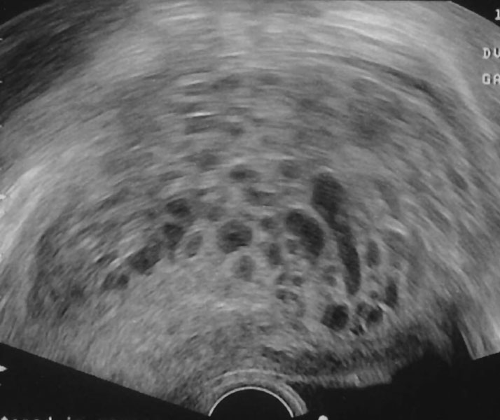
Hydatidiform mole ultrasound
Transvaginal ultrasonography showing a molar pregnancy, the ultrasound pattern is described as a "bunch of grapes".
- Links: Hydatidiform Mole | Ultrasound
Reference
Häggström, Mikael. "Medical gallery of Mikael Häggström 2014". Wikiversity Journal of Medicine 1 (2). DOI:10.15347/wjm/2014.008. ISSN 20018762.
Copyright
This file is made available under the Creative Commons CC0 1.0 Universal Public Domain Dedication. The person who associated a work with this deed has dedicated the work to the public domain by waiving all of his or her rights to the work worldwide under copyright law, including all related and neighboring rights, to the extent allowed by law. You can copy, modify, distribute and perform the work, even for commercial purposes, all without asking permission.
Cite this page: Hill, M.A. (2024, April 19) Embryology Hydatidiform mole ultrasound 01.jpg. Retrieved from https://embryology.med.unsw.edu.au/embryology/index.php/File:Hydatidiform_mole_ultrasound_01.jpg
- © Dr Mark Hill 2024, UNSW Embryology ISBN: 978 0 7334 2609 4 - UNSW CRICOS Provider Code No. 00098G
File history
Click on a date/time to view the file as it appeared at that time.
| Date/Time | Thumbnail | Dimensions | User | Comment | |
|---|---|---|---|---|---|
| current | 10:38, 24 October 2015 |  | 714 × 600 (54 KB) | Z8600021 (talk | contribs) |
You cannot overwrite this file.
File usage
There are no pages that use this file.
