File:Hunter1935 plate01.jpg

Original file (1,790 × 2,107 pixels, file size: 711 KB, MIME type: image/jpeg)
Plate I
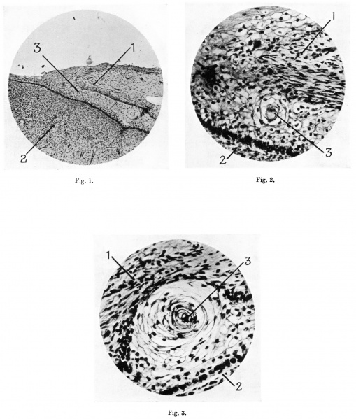
Fig. 1. Microphotograph of terminal part of penis from human foetus of 100 mm. C.R. length, to show the continuity of the cells of the epitriehial layer of the prepuee with those of the layer between the prepuee and glans. Pointers: 1, prepuce; 2, glans penis; 3, cell nest.
Fig. 2. Microphotograph of a “cell-nest” in the layer of cells between prepuce and glans, from a human foetus of 100 mm. C.R. length. Pointers as fig. 1.
Fig. 3. Microphotograph of a “cell-nest” in the layer of cell between prepuce and glans, from a human foetus of 170 mm. C.R.. length. Pointers as fig. 1.
Reference
Hunter RH. Notes on the development of the prepuce. (1935) J Anat. 70: 68-75. PMID 17104576
Cite this page: Hill, M.A. (2024, April 25) Embryology Hunter1935 plate01.jpg. Retrieved from https://embryology.med.unsw.edu.au/embryology/index.php/File:Hunter1935_plate01.jpg
- © Dr Mark Hill 2024, UNSW Embryology ISBN: 978 0 7334 2609 4 - UNSW CRICOS Provider Code No. 00098G
File history
Click on a date/time to view the file as it appeared at that time.
| Date/Time | Thumbnail | Dimensions | User | Comment | |
|---|---|---|---|---|---|
| current | 16:27, 4 January 2017 | | 1,790 × 2,107 (711 KB) | Z8600021 (talk | contribs) | |
| 16:26, 4 January 2017 | 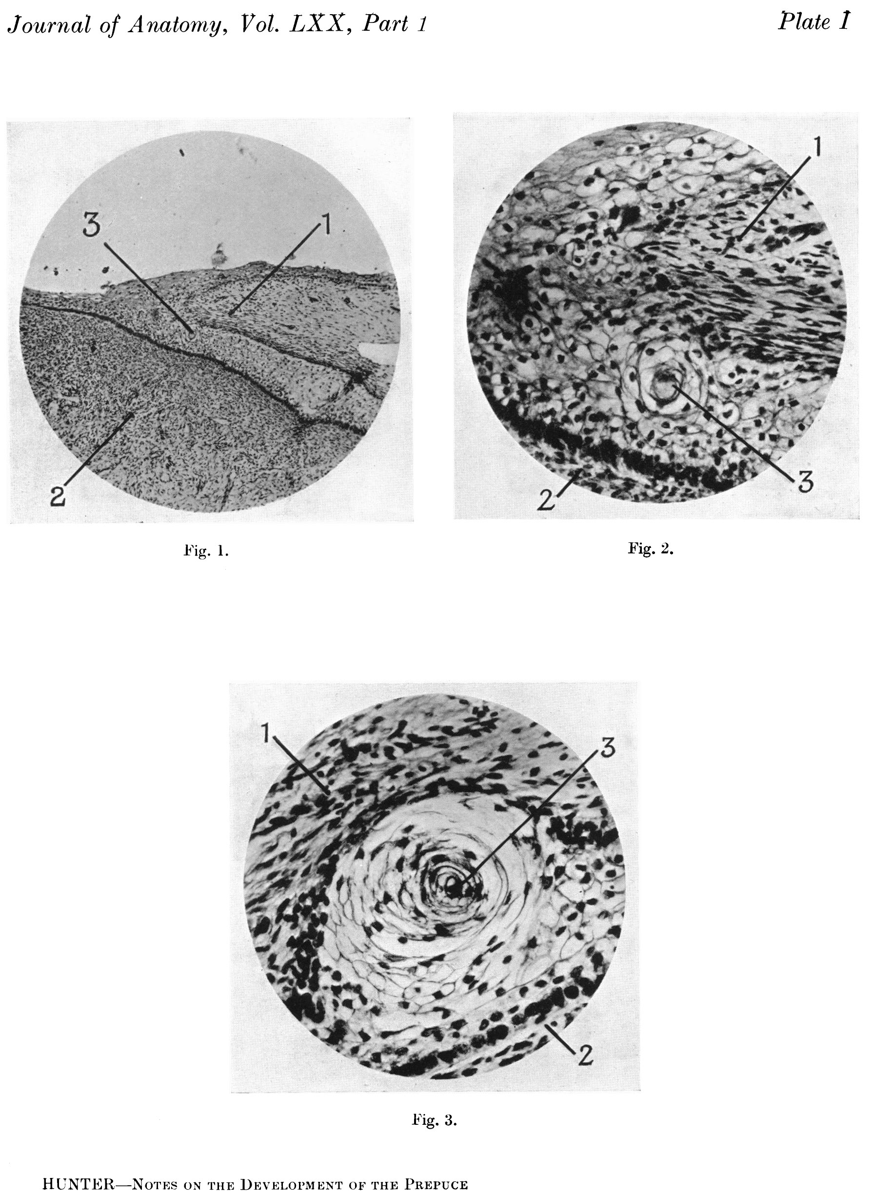 | 1,790 × 2,452 (802 KB) | Z8600021 (talk | contribs) | ==Explanation of Plate I== Fig. 1. Microphotograph of terminal part of penis from human foetus of 100 mm. C.R. length, to show the continuity of the cells of the epitriehial layer of the prepuee with those of the layer between the prepuee and glans. P... |
You cannot overwrite this file.
File usage
The following page uses this file: