File:Human intestine “en-bloc rotation” model.jpg

Original file (1,280 × 635 pixels, file size: 118 KB, MIME type: image/jpeg)
Human intestine “en-bloc rotation” Model
Graphical summary of the “en-bloc rotation” and our hierarchical model of gut morphogenesis. Panels a-c show the classic “en-bloc rotation” model of gut morphogenesis. From panel a to b (left-sided views) the midgut loop rotates 90° in a counterclockwise direction, so that its position changes from midsagittal (a) to transverse (b1). The small intestine forms loops (b2) and slides back into the abdomen (b3) during resolution of the hernia. Meanwhile, the cecum moves from the left to the right side, which represents the additional 180° counterclockwise rotation of the intestine (c, ventral view). The present study shows that the gut acquires its definitive shape by the hierarchical development of primary, secondary, and tertiary loops (panels d-f). The descent of the proximal midgut in the still helically shaped body rather than rotation accounts for the change in position of the primary loop from midsagittal (d, left-sided view) to transverse (e1 left-sided view). During the herniation phase, 4 secondary loops develop in a strict spatiotemporal fashion in the small intestine (e2 left-sided view). Tertiary loops develop within the secondary loops (e3) and these domains slide in a proximodistal fashion back into the abdomen, with the distal ileum and appendix last (e4 left-sided view). Just after return, the cecum is found medially, just dorsal to the umbilical opening. Within 4 days the cecum then assumes its right-sided, more caudal and dorsal position (f1 (colon only), f2 (colon and small intestine); ventral views)
Links: secondary loops week 7 to 8 | tertiary loops week 8 | “en-bloc rotation” model | Intestine Development | Week 8
Reference
<pubmed>26297675</pubmed>
https://bmcdevbiol.biomedcentral.com/articles/10.1186/s12861-015-0081-x
Copyright
© 2015 Soffers et al. Open Access This article is distributed under the terms of the Creative Commons Attribution 4.0 International License (http://creativecommons.org/licenses/by/4.0), which permits unrestricted use, distribution, and reproduction in any medium, provided you give appropriate credit to the original author(s) and the source, provide a link to the Creative Commons license, and indicate if changes were made. The Creative Commons Public Domain Dedication waiver (http://creativecommons.org/publicdomain/zero/1.0/) applies to the data made available in this article, unless otherwise stated.
Fig. 13 Image resized and PMID added.
Cite this page: Hill, M.A. (2024, April 19) Embryology Human intestine “en-bloc rotation” model.jpg. Retrieved from https://embryology.med.unsw.edu.au/embryology/index.php/File:Human_intestine_%E2%80%9Cen-bloc_rotation%E2%80%9D_model.jpg
- © Dr Mark Hill 2024, UNSW Embryology ISBN: 978 0 7334 2609 4 - UNSW CRICOS Provider Code No. 00098G
File history
Click on a date/time to view the file as it appeared at that time.
| Date/Time | Thumbnail | Dimensions | User | Comment | |
|---|---|---|---|---|---|
| current | 13:37, 24 January 2017 | | 1,280 × 635 (118 KB) | Z8600021 (talk | contribs) | |
| 13:35, 24 January 2017 | 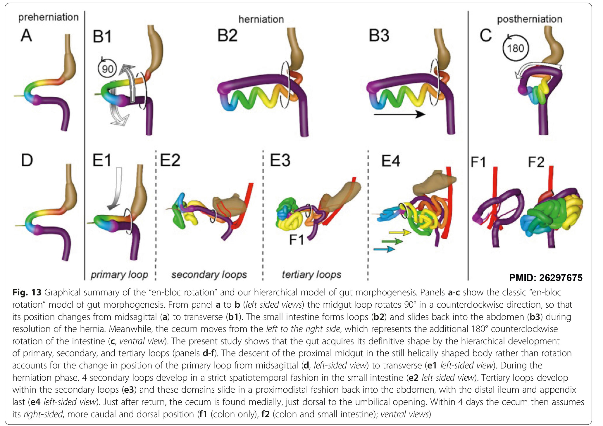 | 2,062 × 1,458 (480 KB) | Z8600021 (talk | contribs) | ==Human intestine “en-bloc rotation” model== ===Reference=== <pubmed>26297675</pubmed> https://bmcdevbiol.biomedcentral.com/articles/10.1186/s12861-015-0081-x ====Copyright==== © 2015 Soffers et al. Open Access This article is distributed under... |
You cannot overwrite this file.
File usage
The following page uses this file: