File:Human infant ovary follicle 01.jpg
From Embryology
Size of this preview: 600 × 600 pixels.
Original file (800 × 800 pixels, file size: 107 KB, MIME type: image/jpeg)
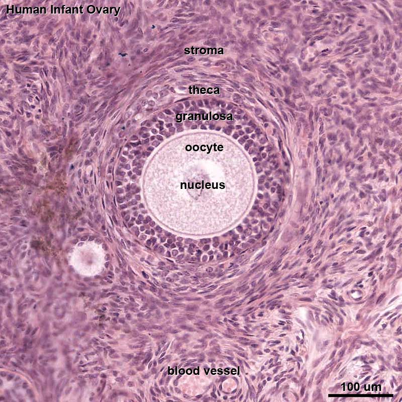
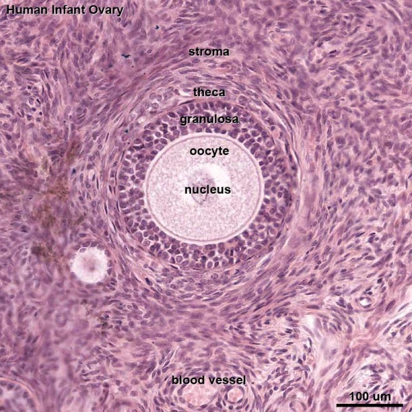
Infant Ovary Follicle (human)
In this infant ovary there are a large number of primordial follicles (oocytes) forming a thick cortical region and no later stages follicle development. Compare this structure with the other images of a mature ovary with reproductive activity.
- primordial follicle - primary oocyte is surrounded by a layer of follicular cells.
- oocytes - enter diplotene stage of prophase.
- Links: Ovary Development | Oocyte Development
Cite this page: Hill, M.A. (2024, April 16) Embryology Human infant ovary follicle 01.jpg. Retrieved from https://embryology.med.unsw.edu.au/embryology/index.php/File:Human_infant_ovary_follicle_01.jpg
- © Dr Mark Hill 2024, UNSW Embryology ISBN: 978 0 7334 2609 4 - UNSW CRICOS Provider Code No. 00098G
File history
Click on a date/time to view the file as it appeared at that time.
| Date/Time | Thumbnail | Dimensions | User | Comment | |
|---|---|---|---|---|---|
| current | 13:37, 5 December 2012 | | 800 × 800 (107 KB) | Z8600021 (talk | contribs) | ==Infant Ovary Follicle (human)== In this infant ovary there are a large number of primordial follicles (oocytes) forming a thick cortical region and no later stages follicle development. Compare this structure with the other images of a mature ovary wit |
You cannot overwrite this file.
File usage
The following page uses this file: