File:Human embryonic shoulder girdle 02.jpg
Original file (1,025 × 713 pixels, file size: 109 KB, MIME type: image/jpeg)
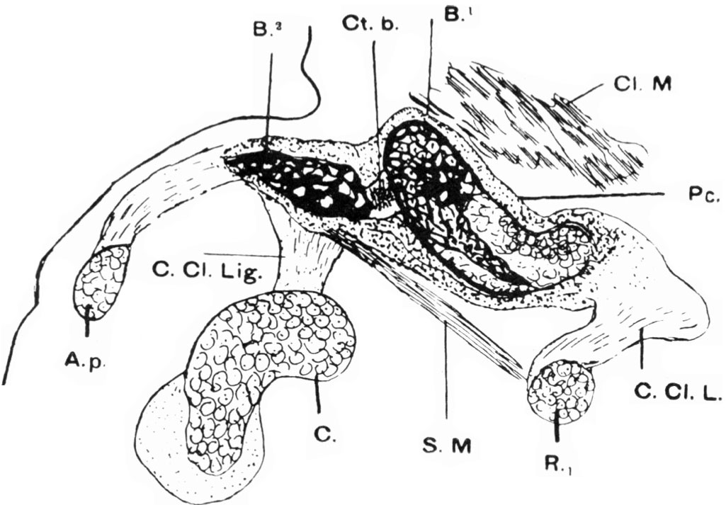
Human Embryonic Shoulder Girdle
Semi-schematic drawing of semi-coronal section of right clavicle of an 18 mm. embryo.
All the parts shown save the subclavius muscle were shown in the section drawn, but the subelavitus was added from its appearance in several sections.
Notice how the sternal segment overrides the acromial segment of the clavicle.
Legend
- A.p - acromion process
- B.1 - bone of sternal segment
- B.2 - bone of acromial segment
- Ct.b. - pre- cartilaginous bridge unossified and connecting acromial and sternal segments
- C. Cl. Lig. - coraco-clavicular ligament
- C - coracoid process
- Cl.M. - cleido-mastoidmuscle
- C.Cl.L. - costo- clavicular ligament
- R.1 - first rib
- S. M. - subclavius muscle
Original file name: FIG.4. Semi-schematic drawing of semi-coronal section of right clavicle of an 18 nm. embryo. (figure scaled and background cleared, above text modified from figure legend)
18mm CRL (Robinson) embryo
- difficult to estimate the amount of shrinkage in this historic material.
- Week 7 Stage 19 CRL 16 - 18 (Somites 48 - 51)
Reference
Fawcett The Development and Ossification of the Human Clavicle. J Anat Physiol: 1913, 47(Pt 2);225-34 PMID 17232952 | PMC1289013
J Anat Physiol. 1913 Jan;47(Pt 2):225-34.
The Development and Ossification of the Human Clavicle.
Fawcett. University of Bristol.
<pubmed>17232952</pubmed>
File history
Click on a date/time to view the file as it appeared at that time.
| Date/Time | Thumbnail | Dimensions | User | Comment | |
|---|---|---|---|---|---|
| current | 09:09, 5 January 2011 | | 1,025 × 713 (109 KB) | S8600021 (talk | contribs) | ==Human Embryonic Shoulder Girdle== Semi-schematic drawing of semi-coronal section of right clavicle of an 18 nm. embryo. All the parts shown save the subclavius muscle were shown in the section drawn, but the subelavitus was added rom its appearance i |
You cannot overwrite this file.
File usage
The following 2 pages use this file: