File:Human embryo femur CS18 to CS23.png

Original file (1,200 × 1,624 pixels, file size: 1.42 MB, MIME type: image/png)
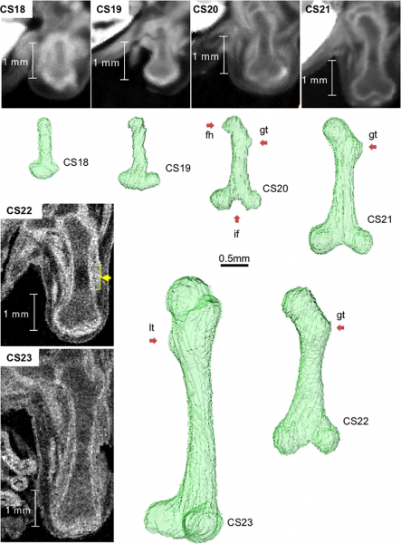
Femur development between CS18 and CS23
(before ossification).
Representative longitudinal section images and 3-D reconstructed images. Longitudinal section images were acquired using PCX-CT between CS18 and CS21 and 7-T MR imaging between CS22 and CS23. fh: femoral head; gt: greater trochanter; If: intercondylar fossa; lt: lesser trochanter; yellow arrow: phase 4 according to Streeter’s classification. See also S1, S2, S3, S4, S5 and S6 Movies.
Reference
Suzuki Y, Matsubayashi J, Ji X, Yamada S, Yoneyama A, Imai H, Matsuda T, Aoyama T & Takakuwa T. (2019). Morphogenesis of the femur at different stages of normal human development. PLoS ONE , 14, e0221569. PMID: 31442281 DOI.
Copyright
© 2019 Suzuki et al. This is an open access article distributed under the terms of the Creative Commons Attribution License, which permits unrestricted use, distribution, and reproduction in any medium, provided the original author and source are credited.
Fig 3. https://doi.org/10.1371/journal.pone.0221569.g003
Cite this page: Hill, M.A. (2024, April 23) Embryology Human embryo femur CS18 to CS23.png. Retrieved from https://embryology.med.unsw.edu.au/embryology/index.php/File:Human_embryo_femur_CS18_to_CS23.png
- © Dr Mark Hill 2024, UNSW Embryology ISBN: 978 0 7334 2609 4 - UNSW CRICOS Provider Code No. 00098G
File history
Click on a date/time to view the file as it appeared at that time.
| Date/Time | Thumbnail | Dimensions | User | Comment | |
|---|---|---|---|---|---|
| current | 21:46, 20 November 2019 |  | 1,200 × 1,624 (1.42 MB) | Z8600021 (talk | contribs) | ==Femur development between CS18 and CS23== (before ossification). Representative longitudinal section images and 3-D reconstructed images. Longitudinal section images were acquired using PCX-CT between CS18 and CS21 and 7-T MR imaging between CS22 a... |
You cannot overwrite this file.
File usage
The following page uses this file: