File:Human and mouse fetal-maternal interface cartoon.jpg
Original file (600 × 720 pixels, file size: 91 KB, MIME type: image/jpeg)
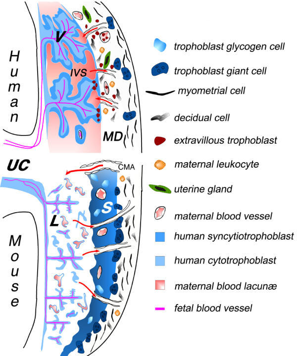
Schematic illustration of the fetal-maternal interface in humans and mice
The placenta, representing the main interface between the mother and fetus, is composed of two parts: the trophoblast of embryonic origin, and the decidua of maternal origin.
During implantation, the trophoblasts derived from the early trophectoderm proliferate rapidly and invade, much like tumor cells, the uterine endometrial tissue. The cell wall of maternal blood vessels encountered by trophoblasts is degraded, causing trophoblasts to be bathed by maternal blood. At the same time, the surrounding maternal tissue is modified extensively, leading to the formation of the decidua. In the human placenta, the syncytiotrophoblast cover of the villi is the main site for all maternofetal transfer and secretory functions, and some of the extravillous cytotrophoblast migrate to an endovascular location, where they can form a new vessel lining, in spiral arteries in particular. Although many differences can be distinguished at the histological level, an increasing number of similarities can be found in the cellular and molecular mechanisms involved in implantation and placental function. Thus, the fetal-maternal interface comprises two main zones of contact, between the fetal trophoblast layers and the maternal decidua, or maternal blood.
Red arrows indicate the blood flow to and from the placenta via maternal arteries or veins, respectively. V: villous trophoblast; IVS: intervillous space; CMA: mouse central maternal artery; S : spongiotrophoblast; L: labyrinthine trophoblast; UC: umbilical cord; MD : maternal decidua.
Reference
<pubmed>14651750</pubmed>| PMC305337 | Reprod Biol Endocrinol.
Copyright
© 2003 Kanellopoulos-Langevin et al; licensee BioMed Central Ltd. This is an Open Access article: verbatim copying and redistribution of this article are permitted in all media for any purpose, provided this notice is preserved along with the article's original URL.
Kanellopoulos-Langevin et al. Reproductive Biology and Endocrinology 2003 1:121 doi:10.1186/1477-7827-1-121
File history
Click on a date/time to view the file as it appeared at that time.
| Date/Time | Thumbnail | Dimensions | User | Comment | |
|---|---|---|---|---|---|
| current | 19:44, 18 May 2013 | | 600 × 720 (91 KB) | Z8600021 (talk | contribs) | Schematic illustration of the fetal-maternal interface in humans and mice. The placenta, representing the main interface between the mother and fetus, is composed of two parts: the trophoblast of embryonic origin, and the decidua of maternal origin. Du... |
You cannot overwrite this file.
File usage
There are no pages that use this file.