File:Human- ventricular system cartoon 02.jpg

Original file (1,179 × 1,254 pixels, file size: 115 KB, MIME type: image/jpeg)
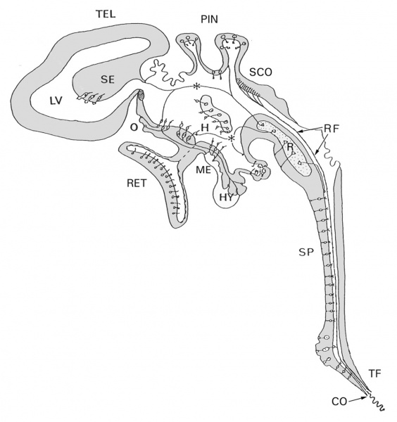
A schematic diagram of structures and specialized cell types bordering the different parts of the mammalian ventricular system, and in contact with the cerebrospinal fluid (CSF).
The complexity of the system suggests that CSF functions are not limited to metabolic support of the brain and the release of waste products.
Abbreviations:
- CO - caudal opening of the central canal of the spinal cord
- H - hypothalamic CSF-contacting neurons
- HY - Hypophysis
- LV - lateral ventricle
- ME - median eminence
- O - vascular organ of the terminal lamina
- PIN - pineal organ
- R - raphe nuclei
- RET - retina
- RF - Reissner's fiber
- SE - septal region
- SCO - subcommissural organ
- SP - medullo-spinal CSF-contacting neurons
- TEL - telencephalon
- TF - terminal filum
Figure. 1 was kindly provided by Prof. B. Vigh to the original article Veening etal., 2010. For details about specific cell types, the reader is referred to: Vigh and Vigh-Teichmann, and to Vigh et al, references listed below.
- Vigh B, Vigh-Teichmann I: Actual problems of the cerebrospinal fluid-contacting neurons. Microsc Res Tech 1998, 41:57-83. Veening and Barendregt Cerebrospinal Fluid Research 2010 7:1 doi:10.1186/1743-8454-7-1
- Vigh B, Manzano e Silva MJ, Frank CL, Vincze C, Czirok SJ, Szabo A, Lukats A, Szel A: The system of cerebrospinal fluid-contacting neurons. Its supposed role in the nonsynaptic signal transmission of the brain.
Reference
Veening JG & Barendregt HP. (2010). The regulation of brain states by neuroactive substances distributed via the cerebrospinal fluid; a review. Cerebrospinal Fluid Res , 7, 1. PMID: 20157443 DOI.
Copyright
© 2010 Veening and Barendregt; licensee BioMed Central Ltd.
This is an Open Access article distributed under the terms of the Creative Commons Attribution License (http://creativecommons.org/licenses/by/2.0), which permits unrestricted use, distribution, and reproduction in any medium, provided the original work is properly cited.
Histol Histopathol 2004, 19:607-628.
Original file name: 1743-8454-7-1-1-l.jpg
Cite this page: Hill, M.A. (2024, April 19) Embryology Human- ventricular system cartoon 02.jpg. Retrieved from https://embryology.med.unsw.edu.au/embryology/index.php/File:Human-_ventricular_system_cartoon_02.jpg
- © Dr Mark Hill 2024, UNSW Embryology ISBN: 978 0 7334 2609 4 - UNSW CRICOS Provider Code No. 00098G
File history
Click on a date/time to view the file as it appeared at that time.
| Date/Time | Thumbnail | Dimensions | User | Comment | |
|---|---|---|---|---|---|
| current | 12:41, 28 April 2010 |  | 1,179 × 1,254 (115 KB) | S8600021 (talk | contribs) | A schematic diagram of structures and specialized cell types bordering the different parts of the mammalian ventricular system, and in contact with the cerebrospinal fluid (CSF). The complexity of the system suggests that CSF functions are not limited t |
You cannot overwrite this file.
File usage
There are no pages that use this file.