File:Horseshoe Kidney.jpg

Original file (787 × 787 pixels, file size: 205 KB, MIME type: image/jpeg)
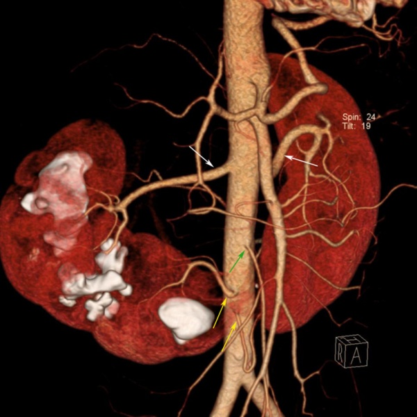
Horseshoe Kidney diagnosis by intravenous pyelography
Diagnosis of horseshoe kidney was made by intravenous pyelography. Coronal volume rendering multidetector CT image shows various blood supply to the horseshoe kidney. Right and left renal arteries (white arrows) supply the upper and middle pole of each kidney, two aortic branches (yellow arrows) supply the lower pole of both kidneys and the isthmus. The isthmus is just below the inferior mesenteric artery (green arrow) origin. Multiple renal stones are also seen in the right kidney.
Reference
<pubmed>22970063</pubmed> Copyright © 2012 Biomedical Imaging and Intervention Journal This is an open-access article distributed under the terms of the Creative Commons Attribution License, which permits unrestricted use, distribution, and reproduction in any medium, provided the original work is properly cited.
--Mark Hill (talk) 15:10 7 November 2014 (EST) Assessment - Figure relates to project topic contains reference, copyright and student template. This abnormality needs to be put in context of fetal renal development.
- Note - This image was originally uploaded as part of an undergraduate science student project and may contain inaccuracies in either description or acknowledgements. Students have been advised in writing concerning the reuse of content and may accidentally have misunderstood the original terms of use. If image reuse on this non-commercial educational site infringes your existing copyright, please contact the site editor for immediate removal.
File history
Click on a date/time to view the file as it appeared at that time.
| Date/Time | Thumbnail | Dimensions | User | Comment | |
|---|---|---|---|---|---|
| current | 09:24, 24 October 2014 |  | 787 × 787 (205 KB) | Z3465141 (talk | contribs) | ==Horseshoe Kidney diagnosis by intravenous pyelography== Diagnosis of horseshoe kidney was made by intravenous pyelography. Coronal volume rendering multidetector CT image shows various blood supply to the horseshoe kidney. Right and left renal arter... |
You cannot overwrite this file.
File usage
The following 2 pages use this file: