File:Holyoke1936 plate 01.jpg

Original file (1,000 × 1,483 pixels, file size: 363 KB, MIME type: image/jpeg)
Plate 1
1 The splenic region of a 6—mm. human enibryo showing the relations in the dorsal mesogastrium before the spleen appears. There is a local bulging of the mesentery but no condensation. The primitive ooelomic epithelium is a dense mantle of cells continuous with the underlying meseneiiynial syneytiuin and with the differeiitiaterl mesothelium at the root of the n1esentery_ Ilemaroxylin and eosin. Photo X 200.
2 The splenic anlage of a 10—mm. pig leml)ry0 showing the dense local mass of cells continuous and morphologically similar h0tl1 with the eoelomic epithelium and the adjacent mesenehyme. Wright’s and Giemsa stain. Photo X 550.
3 The splenie anlage of a 12-mm. pig embryo. The cells are a.1'r2111g'e(l in :1 single layer and a ‘ light zone’ is apparent at some points between the peripheral nuclear stratum and the nleseiiehyme. There is no basement membrane and the cells at a, b and e are niigratirig into the mesenchyme. The apparent double nuclear layer at some points inclioates that the section is not exactly transverse. I)elafield’s hematoxylin and azure ll eosin. Photo X 750.
4 Human embryo, 11 mm, showing the early buckling of the dorsal meso,<2;ast1'ium toward the left and the appearance of the splenic vessels in the coneavity of the resulting fold. The splenie anlage is indieatecl by the prorninenee at the eoxnexity of the fold as it does not stand out clearly from the adjacent dense mesenchyme. The eoelomie epithelium has become a simple eolumnar layer, but there is no basement membrane. There is a. well-defined euboidal. layer on the concave surface of the mesenteric fold. llematoxylin and eosin. ‘Photo X 200.
5 Human embryo, 13.5 mm. The splenie anlage is a distinct condensation of mesenchyme. The overlying eoelomio epithelium has become a low columnar layer marked off by a. basement membrane except in a few places (fig. 6). This columnar layer is continuous with the cuboidal layers beyond the margins of the anlage. Hematoxylin and eosin. Photo X 200.
6 Detail from preceding figure. The (tell me. is being added to the splenio Inesenchyme. nm. is a typical mesenchymal nueleus in one of the epithelial cells. Photo X 400.
| Historic Disclaimer - information about historic embryology pages |
|---|
 |
Reference
Holyoke EA. The role of the primitive mesothelium in the development of the mammalian spleen. (1936) Anat. Rec. 65(3): 333-349.
Cite this page: Hill, M.A. (2024, April 19) Embryology Holyoke1936 plate 01.jpg. Retrieved from https://embryology.med.unsw.edu.au/embryology/index.php/File:Holyoke1936_plate_01.jpg
- © Dr Mark Hill 2024, UNSW Embryology ISBN: 978 0 7334 2609 4 - UNSW CRICOS Provider Code No. 00098G
File history
Click on a date/time to view the file as it appeared at that time.
| Date/Time | Thumbnail | Dimensions | User | Comment | |
|---|---|---|---|---|---|
| current | 16:55, 20 March 2017 | | 1,000 × 1,483 (363 KB) | Z8600021 (talk | contribs) | |
| 16:55, 20 March 2017 | 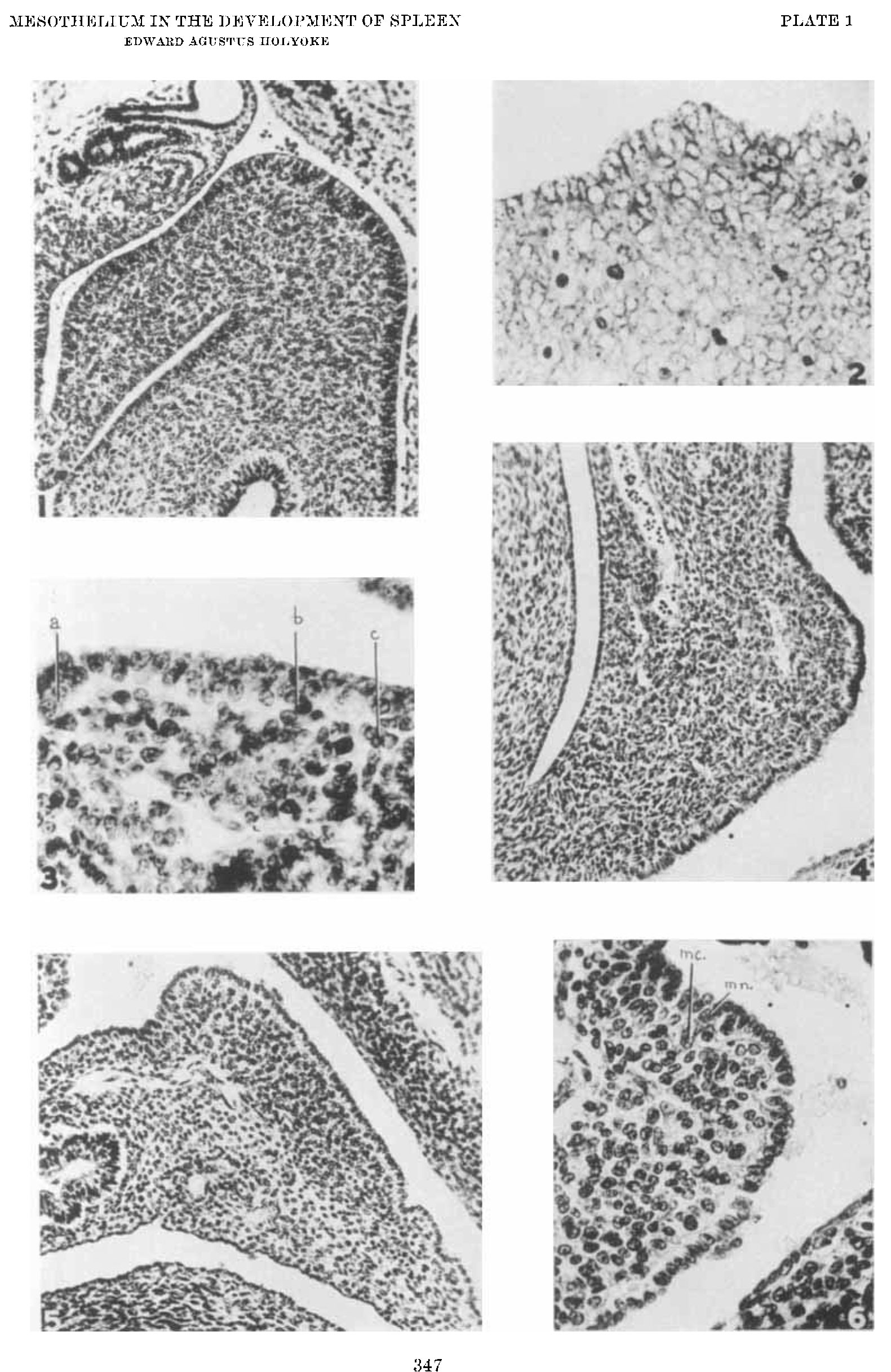 | 1,581 × 2,434 (565 KB) | Z8600021 (talk | contribs) | {{Historic Disclaimer}} ===Reference=== {{Ref-Holyoke1936}} {{Footer}} Category:Spleen |
You cannot overwrite this file.
File usage
The following page uses this file: