File:Histology terminology cartoon.jpg
From Embryology
Size of this preview: 423 × 599 pixels. Other resolution: 595 × 842 pixels.
Original file (595 × 842 pixels, file size: 72 KB, MIME type: image/jpeg)
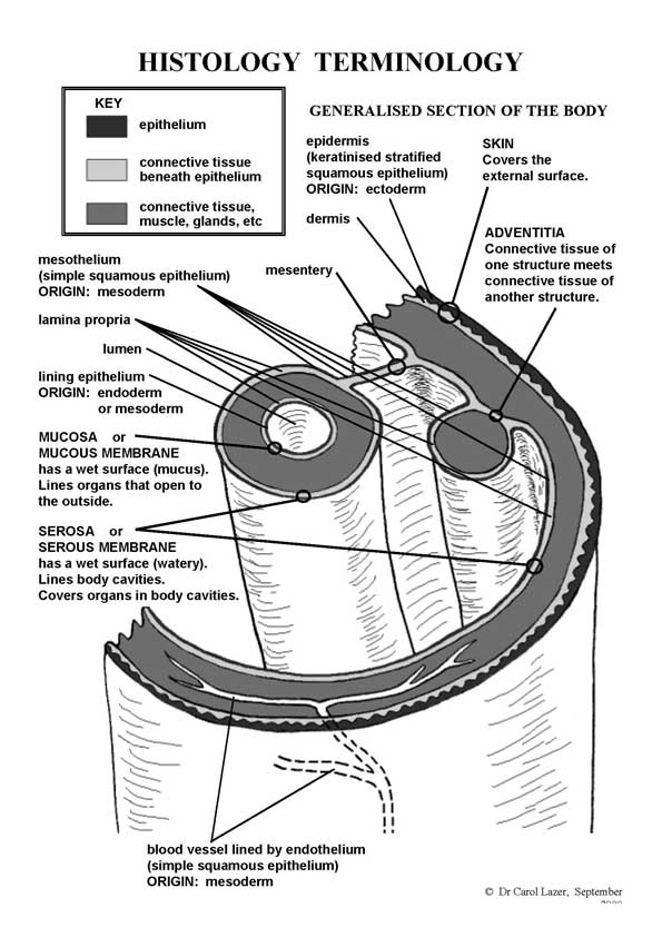
Histology Terminology
Skin
Covers the external surface.
- epidermis - epithelium, keratinised stratified squamous epithelium.
- dermis - connective tissue; papillary (loose CT) and reticular layer (dense irregular CT)
- hypodermis - (hypoderm, subcutis) connective tissue,
Adventitia
Within the body.
- Connective tissue of one structure meets connective tissue of another structure.
Serosa
(serous membrane) has a wet surface (watery).
- Epithelia and connective tissue lines body cavities. Covers organs in the body.
- mesothelium - the layer of cells (mesoderm in origin), lining the body cavity of the embryo. In the adult, it is a simple squamous epithelium that covers all true serous membranes (peritoneum, pericardium, pleura).
Mucosa
(mucous membrane) has a wet surface (mucus).
- Epithelia and connective tissue that lines organs that open to the outside.
- Links: Foundations - Histology Cells and Tissues | Foundations - Histology Epithelia and Skin | Image - Skin structure | Histology | Integumentary System Development
Reference
Diagram prepared by Dr Carol Lazar.
File history
Click on a date/time to view the file as it appeared at that time.
| Date/Time | Thumbnail | Dimensions | User | Comment | |
|---|---|---|---|---|---|
| current | 16:00, 14 February 2012 | | 595 × 842 (72 KB) | S8600021 (talk | contribs) | ==Histology terminology cartoon== Diagram prepared by Dr Carol Lazar. Category:Histology |
You cannot overwrite this file.