File:His1897 fig07.jpg
From Embryology
Size of this preview: 799 × 180 pixels. Other resolution: 1,584 × 357 pixels.
Original file (1,584 × 357 pixels, file size: 68 KB, MIME type: image/jpeg)
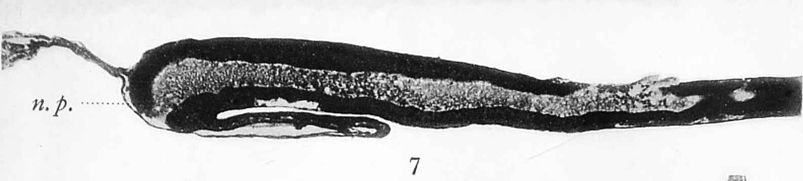
Fig 7. Mesial longitudinal section through the neural tube of an early chick embryo
The aperture or neuropore (n.p.) at the anterior end of the tube is still open.
| Historic Disclaimer - information about historic embryology pages |
|---|
 |
- Links: Fig 1 | Fig 2 | Fig 3 | Fig 4 | Fig 5 | Fig 6 | Plate 1 | Fig 7 | Fig 8 | Fig 9 | Fig 10 | Fig 11 | Plate 2 | Fig 12 | Fig 13 | Fig 14 | Fig 15 | Plate 3 | Fig 16 | Fig 17 | Plate 4 | Fig 18 | Fig 19 | Fig 20 | Fig 21 | Fig 22 | Fig 23 | Plate 5 | Fig 24 | Fig 25 | Fig 26 | Fig 27 | Fig 28 | His 1897 | Wilhelm His | Historic Embryology Papers
Reference
His W. Address upon the development of the brain. (1897) Trans. Royal Acad. Medicine Ireland.
Cite this page: Hill, M.A. (2024, April 16) Embryology His1897 fig07.jpg. Retrieved from https://embryology.med.unsw.edu.au/embryology/index.php/File:His1897_fig07.jpg
- © Dr Mark Hill 2024, UNSW Embryology ISBN: 978 0 7334 2609 4 - UNSW CRICOS Provider Code No. 00098G
File history
Click on a date/time to view the file as it appeared at that time.
| Date/Time | Thumbnail | Dimensions | User | Comment | |
|---|---|---|---|---|---|
| current | 14:44, 18 January 2016 | 1,584 × 357 (68 KB) | Z8600021 (talk | contribs) | ===Plate I=== Mesial longitudinal section through the neural tube of an early chick embryo. The aperture or neuropore (n.p.) at the anterior end of the tube is still open. Fig. 8. Mesial longitudinal section through the head-end of a slightly older ch... |
You cannot overwrite this file.
File usage
The following 2 pages use this file: