File:Hertwig1892 plate2.jpg

Original file (2,295 × 1,400 pixels, file size: 667 KB, MIME type: image/jpeg)
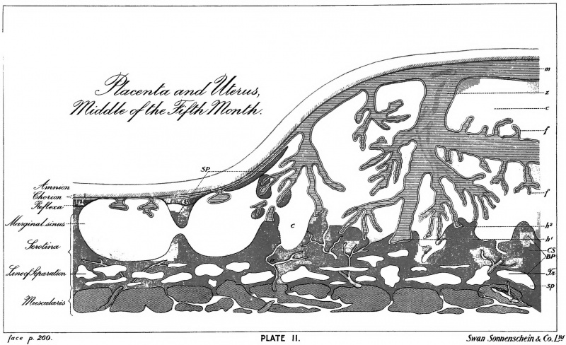
Plate II. Diagrammatic section through the human placenta at the middle of the Fifth month
after LEOPOLD.
The musculature of the uterus is followed by the spongy layer of the clecidua serotina (sp), in which the separation of the placenta takes place at birth along the line of separation indicated by two heavy marks ; this is followed by the compact layer ( CS), which is thrown off at birth as the placenta uterina, and which consists of the (WINKLER'S) basal plate (BP), closing plate (Schlussplatte) (P), cavernous blood-spaces (>), the arteria advehentes (), and the marginal sinus. The placenta foetalis has grown into the placenta uterina; it consists of the membrana chorii (m~) and the villi (z) arising- from it ; on the latter are to be distinguished the roots of attachment (h\ /j, 2 ) and the free processes (/). [ejj, Foetal epithelium derived from the serosa.] The chorion is still covered internally by the amnion. (The foetal part of the placenta is reproduced in blue, the maternal part in black and brown ; pink indicates the blood-spaces.)
File history
Click on a date/time to view the file as it appeared at that time.
| Date/Time | Thumbnail | Dimensions | User | Comment | |
|---|---|---|---|---|---|
| current | 16:01, 21 February 2015 | | 2,295 × 1,400 (667 KB) | Z8600021 (talk | contribs) |
You cannot overwrite this file.
File usage
The following page uses this file: