File:Hertwig1892 fig323.jpg
From Embryology

Size of this preview: 687 × 599 pixels. Other resolution: 729 × 636 pixels.
Original file (729 × 636 pixels, file size: 132 KB, MIME type: image/jpeg)
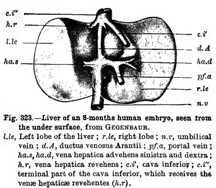
Fig. 323. Liver of an 8-months human embryo
Seen from the under surface, from GKGENBAUR. Lie, Left lobe of the liver ; r.le, right lobe ; n.r, umbilical vein ; d.A, ductus venosus Arantii ; pf.a, portal vein ; ha. s, ha.d, vena hepatica advehens sinistra and dextra ; h.r, vena hepatica revehens ; c.i', cava inferior; c.i", terminal part of the cava inferior, which receives the vente hepaticae revehentes (h.r.).
| Historic Disclaimer - information about historic embryology pages |
|---|
| Pages where the terms "Historic" (textbooks, papers, people, recommendations) appear on this site, and sections within pages where this disclaimer appears, indicate that the content and scientific understanding are specific to the time of publication. This means that while some scientific descriptions are still accurate, the terminology and interpretation of the developmental mechanisms reflect the understanding at the time of original publication and those of the preceding periods, these terms, interpretations and recommendations may not reflect our current scientific understanding. (More? Embryology History | Historic Embryology Papers) |
Reference
Historic Textbook Text-Book of the Embryology of Man and Mammals by Dr Oscar Hertwig (1892)
File history
Click on a date/time to view the file as it appeared at that time.
| Date/Time | Thumbnail | Dimensions | User | Comment | |
|---|---|---|---|---|---|
| current | 15:46, 21 February 2015 |  | 729 × 636 (132 KB) | Z8600021 (talk | contribs) |
You cannot overwrite this file.
File usage
The following page uses this file:
