File:Hertig1946b fig25b.jpg
Original file (800 × 644 pixels, file size: 139 KB, MIME type: image/jpeg)
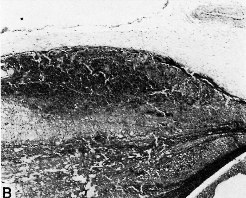
Fig. 25B. A human placenta at term
To show diffuse and discrete thrombosis of intervillous space. B. L.—i. H., S-32-217.
A more highly magnified view of rectangle seen in 25 A. The fibrous tissue of the chorionic membrane is at the top, beneath which is the cytotrophoblast whose “fibrinoid” degeneration has caused the discrete mass of fibrin to be deposited from the maternal blood. X60.
References
Hertig AT. lnvolution of tissues in fetal life: a review. (1946) Anat. Rec. 94: 96-116.
Cite this page: Hill, M.A. (2024, April 20) Embryology Hertig1946b fig25b.jpg. Retrieved from https://embryology.med.unsw.edu.au/embryology/index.php/File:Hertig1946b_fig25b.jpg
- © Dr Mark Hill 2024, UNSW Embryology ISBN: 978 0 7334 2609 4 - UNSW CRICOS Provider Code No. 00098G
File history
Click on a date/time to view the file as it appeared at that time.
| Date/Time | Thumbnail | Dimensions | User | Comment | |
|---|---|---|---|---|---|
| current | 13:05, 8 August 2017 | | 800 × 644 (139 KB) | Z8600021 (talk | contribs) | ==Fig. 25. A human placenta at term== To show diffuse and discrete thrombosis of intervillous space. B. L.—i. H., S-32-217. A. Laminated fibrin thrombus deposited on under surface of chorionic membrane at left of picture. Note septum running horiz... |
You cannot overwrite this file.
File usage
The following page uses this file: