File:Hertig1946b fig07a.jpg
Hertig1946b_fig07a.jpg (800 × 569 pixels, file size: 80 KB, MIME type: image/jpeg)
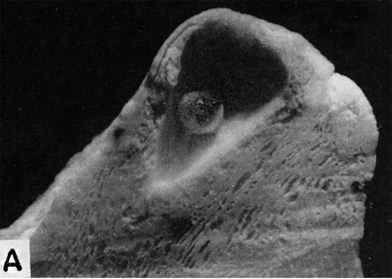
Fig. 7A. A human embryo of approximately 20 - 21 days of age
A human embryo of approximately 20 - 21 days of age, measuring 1.6 mm in length. Fig. A. represents the chorion embedded within the endometrium and opened in such a way as to reveal the entire embryo when viewed from its left side. The yolk-sac is the most prominent feature of the embryo and is seen as a balloon-like mass whose distal surface is covered with blood islands. The embryonic disk itself together with its amnion is the thickened opaque mass where the embryo is attached to the chorion. See fig. B. for morphologic details of the embryo. Carnegie 7545, sequence 7, X5.
References
Hertig AT. lnvolution of tissues in fetal life: a review. (1946) Anat. Rec. 94: 96-116.
Cite this page: Hill, M.A. (2024, April 19) Embryology Hertig1946b fig07a.jpg. Retrieved from https://embryology.med.unsw.edu.au/embryology/index.php/File:Hertig1946b_fig07a.jpg
- © Dr Mark Hill 2024, UNSW Embryology ISBN: 978 0 7334 2609 4 - UNSW CRICOS Provider Code No. 00098G
File history
Click on a date/time to view the file as it appeared at that time.
| Date/Time | Thumbnail | Dimensions | User | Comment | |
|---|---|---|---|---|---|
| current | 17:09, 7 August 2017 |  | 800 × 569 (80 KB) | Z8600021 (talk | contribs) |
You cannot overwrite this file.
File usage
The following page uses this file:
