File:Hertig1946b fig06.jpg
Hertig1946b_fig06.jpg (800 × 582 pixels, file size: 132 KB, MIME type: image/jpeg)
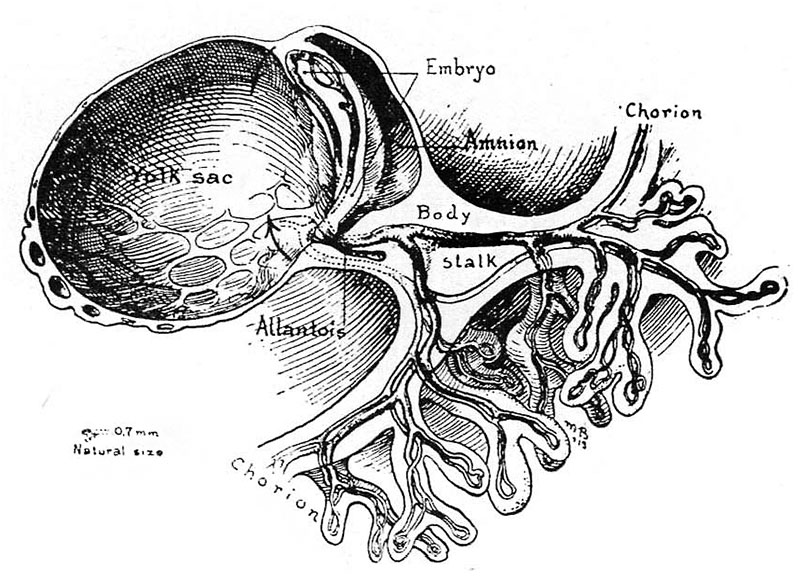
Fig 6. A drawing of a 0.7 mm human embryo, about 17 days of age
Taken from Cullen’s “The Umbilicus and Its Diseases,” W. B. Saunders Company. This figure, depicting the right half of the embryonic mass shows the circulation in the yolksac, embryo, body‘stalk and chorion as anatomically complete although later studies show that such a state does not actually occur until about 3 days later. Note the relatively enormous size of the yolk-sac with respect to the embryo and its amnion. Only the portion of the chorion or outer shell of the ovum near the body stalk is represented, the entire chorion at this stage measuring about 8.0 mm. in diameter.
References
Hertig AT. lnvolution of tissues in fetal life: a review. (1946) Anat. Rec. 94: 96-116.
Cite this page: Hill, M.A. (2024, April 18) Embryology Hertig1946b fig06.jpg. Retrieved from https://embryology.med.unsw.edu.au/embryology/index.php/File:Hertig1946b_fig06.jpg
- © Dr Mark Hill 2024, UNSW Embryology ISBN: 978 0 7334 2609 4 - UNSW CRICOS Provider Code No. 00098G
File history
Click on a date/time to view the file as it appeared at that time.
| Date/Time | Thumbnail | Dimensions | User | Comment | |
|---|---|---|---|---|---|
| current | 17:05, 7 August 2017 | | 800 × 582 (132 KB) | Z8600021 (talk | contribs) |
You cannot overwrite this file.
File usage
The following page uses this file: