File:Hertig1945d fig10.jpg
Original file (1,000 × 833 pixels, file size: 151 KB, MIME type: image/jpeg)
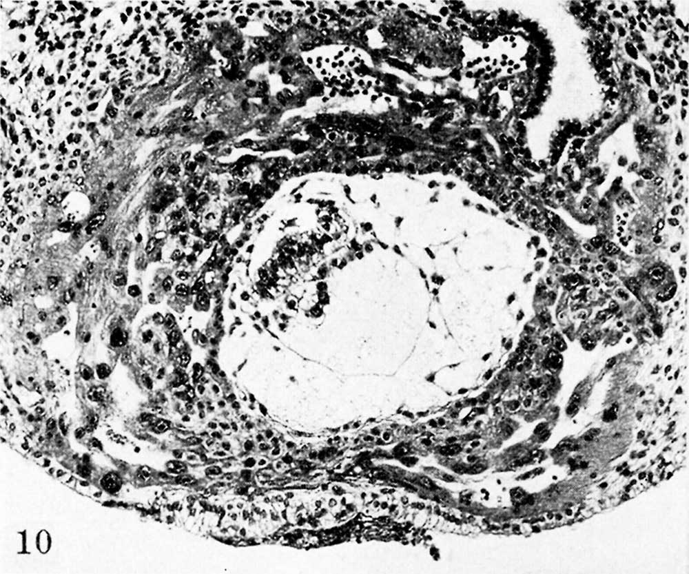
Fig. 10. A mid-cross section of an 11.5-day ovum
The amniotic cavity is more advanced in its development than in the previous specimens although the amniogenic cells— now a distinct membrane - are still attached to the adjacent trophoblast. The exocoelome is now well contained within its membrane which is continuous above with the primitive endoderm of the germ-disk. Mesoblast formation is still continuing and is laying the foundation for the future connective tissue of the chorion. Carnegie 7699 section 8-5-3, X 100.
Reference
Hertig AT. On the development of the amnion and exocoelomic membrane in the previllous human ovum. (1945) Yale J Biol Med. 18:107-15. PubMed 21007544
Cite this page: Hill, M.A. (2024, April 25) Embryology Hertig1945d fig10.jpg. Retrieved from https://embryology.med.unsw.edu.au/embryology/index.php/File:Hertig1945d_fig10.jpg
- © Dr Mark Hill 2024, UNSW Embryology ISBN: 978 0 7334 2609 4 - UNSW CRICOS Provider Code No. 00098G
File history
Click on a date/time to view the file as it appeared at that time.
| Date/Time | Thumbnail | Dimensions | User | Comment | |
|---|---|---|---|---|---|
| current | 15:49, 24 October 2017 | | 1,000 × 833 (151 KB) | Z8600021 (talk | contribs) |
You cannot overwrite this file.