File:Hertig1945d fig07.jpg
Original file (1,280 × 601 pixels, file size: 138 KB, MIME type: image/jpeg)
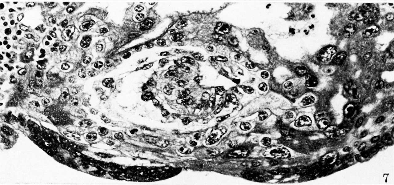
Fig. 7. A mid-cross section of an ovum 8 to 9 days of age
Note that the amniotic cavity is no more advanced than is that of the younger specimen seen in ii . 6, although there are several amniogenic cells arising from the adjacent trophoblast in the ol er specimen. Likewise, mesoblastic formation is more advanced, as shown by the presence of an early exocoelomic membrane. In places the mesoblastic cells of the latter are still attached to the trophoblast from which they appear to be originating. Carnegie 8171, section 3-2-12, X 300.


Reference
Hertig AT. On the development of the amnion and exocoelomic membrane in the previllous human ovum. (1945) Yale J Biol Med. 18:107-15. PubMed 21007544
Cite this page: Hill, M.A. (2024, April 24) Embryology Hertig1945d fig07.jpg. Retrieved from https://embryology.med.unsw.edu.au/embryology/index.php/File:Hertig1945d_fig07.jpg
- © Dr Mark Hill 2024, UNSW Embryology ISBN: 978 0 7334 2609 4 - UNSW CRICOS Provider Code No. 00098G
File history
Click on a date/time to view the file as it appeared at that time.
| Date/Time | Thumbnail | Dimensions | User | Comment | |
|---|---|---|---|---|---|
| current | 15:37, 24 October 2017 | | 1,280 × 601 (138 KB) | Z8600021 (talk | contribs) |
You cannot overwrite this file.