File:Herring1908b fig07.jpg
Original file (1,280 × 974 pixels, file size: 290 KB, MIME type: image/jpeg)
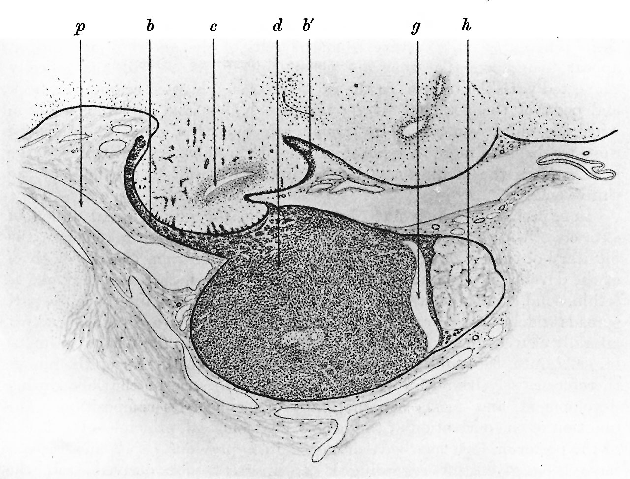
Fig. 7. Pituitary body of a human foetus (fifth month)
Sagittal section through same pituitary as shown in fig. 6, but further to one side. Drawing from a photograph.
b, tongue-like process of epithelium spreading forward: b’, epithelial cells spreading backwards over surface of the brain ; b, third ventricle ; d, anterior lobe ; g, epithelial cleft; h, posterior lobe; p, lymph space.
Reference
Herring PT. The development of the mammalian pituitary and its morphological significance. (1908) Quar. Jour. Ex. Physiol. 1: 161-185.
Cite this page: Hill, M.A. (2024, April 25) Embryology Herring1908b fig07.jpg. Retrieved from https://embryology.med.unsw.edu.au/embryology/index.php/File:Herring1908b_fig07.jpg
- © Dr Mark Hill 2024, UNSW Embryology ISBN: 978 0 7334 2609 4 - UNSW CRICOS Provider Code No. 00098G
File history
Click on a date/time to view the file as it appeared at that time.
| Date/Time | Thumbnail | Dimensions | User | Comment | |
|---|---|---|---|---|---|
| current | 13:12, 6 September 2018 | | 1,280 × 974 (290 KB) | Z8600021 (talk | contribs) | |
| 13:12, 6 September 2018 |  | 1,556 × 1,317 (612 KB) | Z8600021 (talk | contribs) | ==Fig. 7. == ===Reference=== {{Ref-Herring1908b}} {{Footer}} Category:HumanCategory:Second TrimesterCategory:Pituitary |
You cannot overwrite this file.
File usage
The following 2 pages use this file: