File:Heartfibertracttractography.png

Original file (2,004 × 1,284 pixels, file size: 3.98 MB, MIME type: image/png)
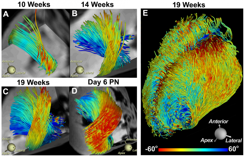
Myofiber tractography of the lateral wall of the left ventricle at different weeks of a human fetal heart.
(A) A few fiber tracts present at week 10. (B) An increase in the density of the fiber tracts and crossing-helical pattern is visible at week 14. (C) At week 19, the arrangement of the fiber tracts is similar to that of the heart after birth. (D) Fiber tracts of an adult heart (PN = P6 neonatal). (E) A complete tractography of the heart at week 19.
Reference [1]
- ↑ <pubmed>25031361</pubmed>
Copyright: © 2013 Mekkaoui et al. This is an open-access article distributed under the terms of the Creative Commons Attribution License, which permits unrestricted use, distribution, and reproduction in any medium, provided the original author and source are credited.
- Note - This image was originally uploaded as part of an undergraduate science student project and may contain inaccuracies in either description or acknowledgements. Students have been advised in writing concerning the reuse of content and may accidentally have misunderstood the original terms of use. If image reuse on this non-commercial educational site infringes your existing copyright, please contact the site editor for immediate removal.
--Mark Hill All the information is here, with some minor format issues. You should have used a file name that describes the image not File:Heartfibertracttractography.png which is not identifiable. (4/5)
File history
Click on a date/time to view the file as it appeared at that time.
| Date/Time | Thumbnail | Dimensions | User | Comment | |
|---|---|---|---|---|---|
| current | 22:57, 18 August 2014 |  | 2,004 × 1,284 (3.98 MB) | Z3417843 (talk | contribs) |
You cannot overwrite this file.
File usage
The following page uses this file: