File:Hassall1849 plate77.jpg

Original file (1,280 × 2,045 pixels, file size: 741 KB, MIME type: image/jpeg)
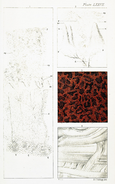
Plate LXXVII.
| Fig. 1. represents a magnified view of a vertical section of the skin
under a power of seventy or eighty diameters: g. g. Sudoriparous glands imbedded in fat vesicles ; d. the ducts of the same passing in a flexuous course through the areolar tissue to de, the dermic portion of the skin ; two of these ducts are represented cut across. On the right, a duct is represented cut open at its upper part, and its parietes are seen to be continuous with the basement membrane of the papillae which bound it on each side, assuming as it approaches them an infundibular form. Between the same two papillae may be seen the lowest portion of the epidermic part of a duct, at first very indistinctly, and without any defined continuity of structure with the duct below — gradually assuming a spiral form, and having the scales of which its walls are composed, arranged parallel with the axis of the passage. The other ducts are seen dipping down between and behind the papillae ; at n, may be seen the nuclei on the basement membrane of the papillae, which at nc are developed into a layer of nucleated cells, forming the lower stratum of the epidermis, ep, through which one complete sudoriferous passage, p, may be seen passing to the surface, together with portions of others. The spaces between these passages have been cut away in the preparation, by which the direction of the scales of the epidermis not in the vicinity of a passage are seen to be horizontal, but variously inclined where situated in its vicinity. After Rainey and Ralph. |
Fig. 2. is a magnified view (220 diameters) of the dermic part; d, the
dermic portion of a duct cut open at its upper part, also with the basement membrane of the papillae on each side continuous with it; p, the epidermic portion of the duct between the papillae, exhibiting a scaly structure almost at its commencement; n, nuclei on the basement membrane, at nc, developed into nucleated cells, and forming together the lower part of the epidermis ; above which, at ep, may be seen the commencement of the scaly layer of the epidermis ; three papillae with a vascular loop in each. After Rainey and Ralph. Fig. 3. Mucous membrane of the gall-bladder ; from an injection by Dr. Jno. Neill, of Philadelphia (see page 358). 50 diameters. Fig. 4. Transverse section of the muscles of the tongue. The fibres are of the striped variety, but are not here sufficiently magnified to show the lines. 45 diameters. |
Reference
Hassall AH. The microscopic anatomy of the human body, in health and disease. (1849) Samuel Hurley, Fleet Street, London.
Cite this page: Hill, M.A. (2024, April 19) Embryology Hassall1849 plate77.jpg. Retrieved from https://embryology.med.unsw.edu.au/embryology/index.php/File:Hassall1849_plate77.jpg
- © Dr Mark Hill 2024, UNSW Embryology ISBN: 978 0 7334 2609 4 - UNSW CRICOS Provider Code No. 00098G
File history
Click on a date/time to view the file as it appeared at that time.
| Date/Time | Thumbnail | Dimensions | User | Comment | |
|---|---|---|---|---|---|
| current | 09:45, 25 January 2019 | | 1,280 × 2,045 (741 KB) | Z8600021 (talk | contribs) | |
| 09:44, 25 January 2019 | 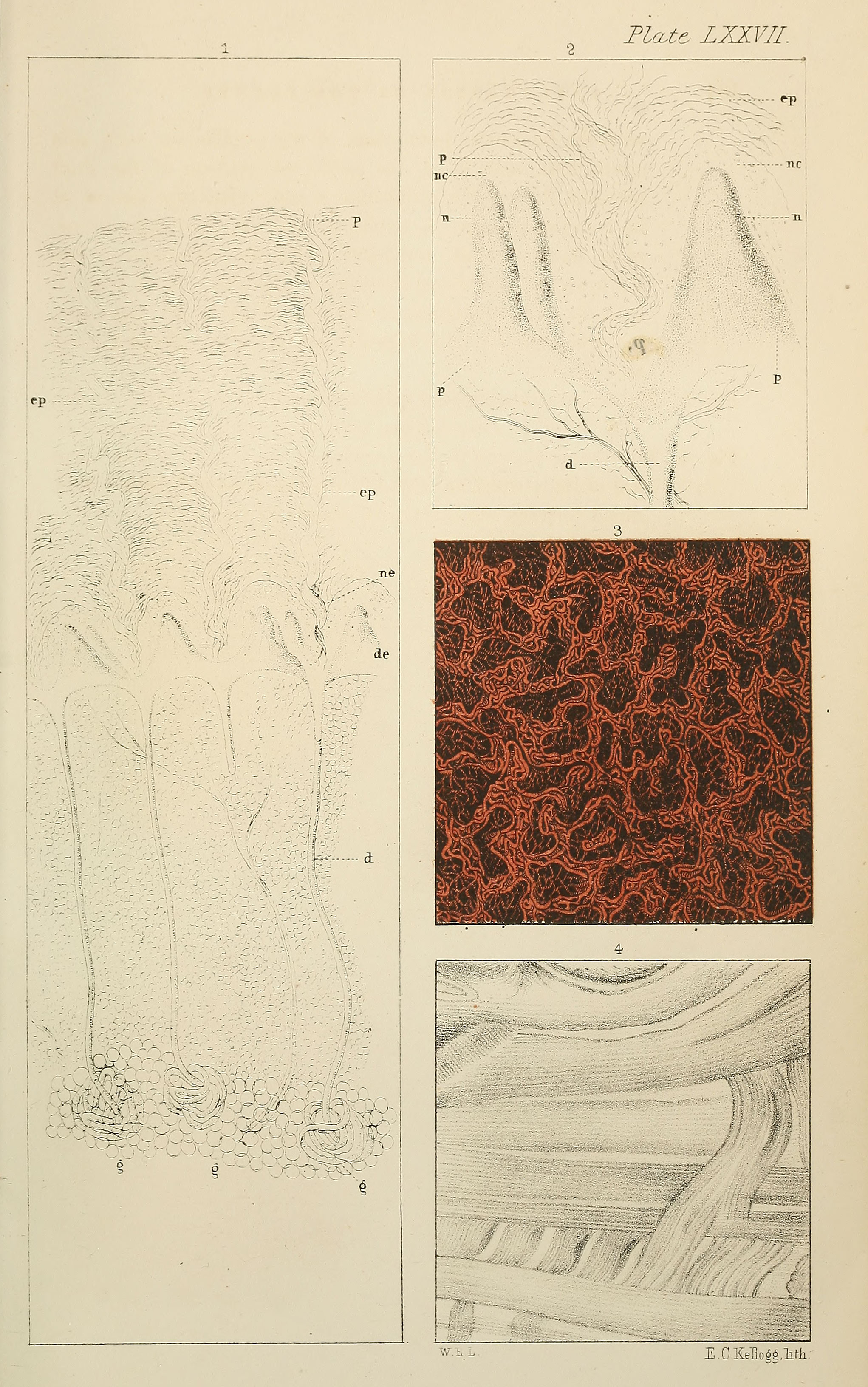 | 2,043 × 3,264 (1.26 MB) | Z8600021 (talk | contribs) | ||
| 09:42, 25 January 2019 | 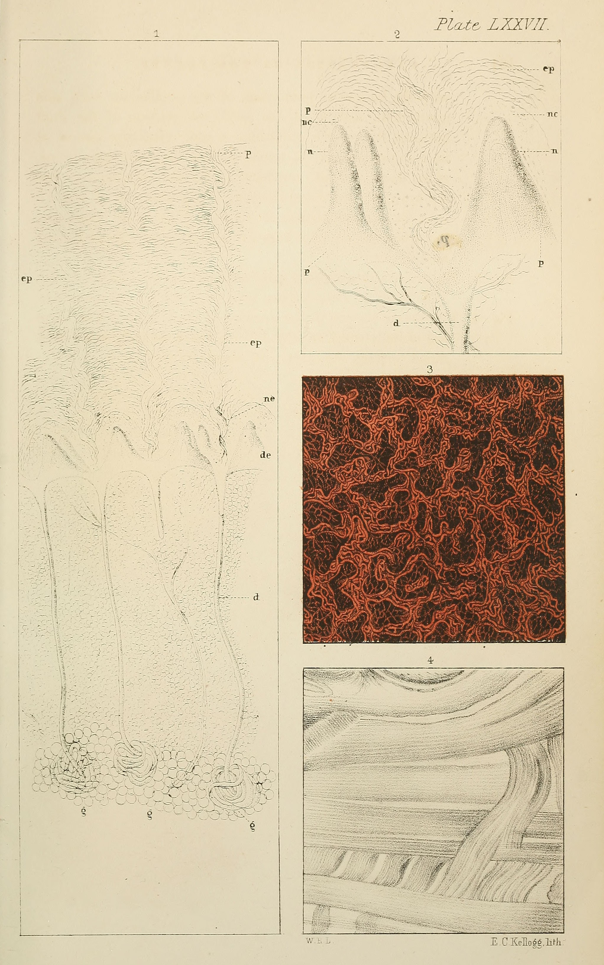 | 2,043 × 3,264 (1.16 MB) | Z8600021 (talk | contribs) |
You cannot overwrite this file.
File usage
The following page uses this file: