File:Hamster oocyte zona pellucida SEM.jpg

Original file (800 × 626 pixels, file size: 101 KB, MIME type: image/jpeg)
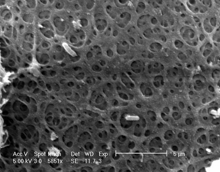
Hamster Oocyte Zona Pellucida SEM
Surface of the zona pellucida surrounding a hamster oocyte. This scanning electron micrograph shows the zona pellicida around a hamster oocyte at high magnification. The zona pellicida has a complex weave and is comprised of three glycoproteins, ZP1, ZP2 and ZP3.
Magnification: 5851x. (Stain - Osmium)
Original image name: 12624.jpg http://www.cellimagelibrary.org/images/12624
The sample was fixed using glutaraldehyde and osmium tetroxide, dehydrated in ethanol, critically point dried, coated with gold, and examined in a Phillips XL30 FEG scanning electron microscope.
Public Domain: This image is in the public domain and thus free of any copyright restrictions. However, as is the norm in scientific publishing and as a matter of courtesy, any user should credit the content provider for any public or private use of this image whenever possible.
File history
Click on a date/time to view the file as it appeared at that time.
| Date/Time | Thumbnail | Dimensions | User | Comment | |
|---|---|---|---|---|---|
| current | 08:12, 26 April 2011 |  | 800 × 626 (101 KB) | S8600021 (talk | contribs) | |
| 08:10, 26 April 2011 |  | 800 × 626 (90 KB) | S8600021 (talk | contribs) | ==Hamster Oocyte Zona Pellucida SEM== Surface of the zona pellucida surrounding a hamster oocyte. This scanning electron micrograph shows the zona pellicida around a hamster oocyte at high magnification. The zona pellicida has a complex weave and is comp |
You cannot overwrite this file.
File usage
The following 3 pages use this file: