File:HamiltonBoyd1960 plate07.jpg
Original file (1,280 × 1,536 pixels, file size: 608 KB, MIME type: image/jpeg)
Plate 7
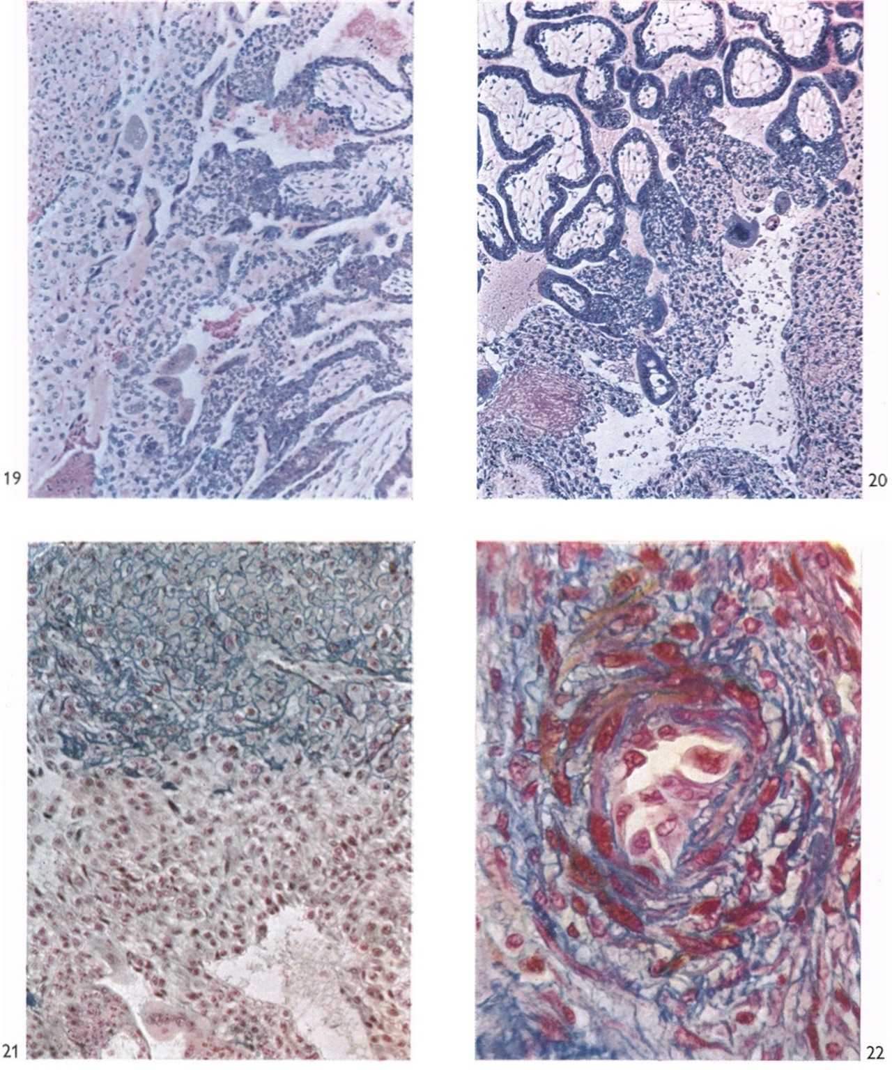
Fig. 19. Photomicrograph (x 100) of a section (H. & E.) of an 18-day embryo (Gar). Marked proliferation at the tips of the villi (to the right) has resulted in the production of a trophoblastic shell which now extends completely around the foetal tissue. The villi are covered except. at their tips by a Langhan’s layer of cytotrophoblast and a superficial layer of definitive syncytium. At one point a syncytial sprout can be seen arising from the definitive syncytium. Remnants of the primitive syncytium lie within and on the maternal aspect of the trophoblastic shell. Early vasculogenesis is present in the mesodermal cores of the villi.
Fig. 20. Photomicrograph ( x 60) of a section (H. & E.) through the tips of the chorionic villi and trophoblastic shell of a 26-day human implantation site (Camb. H. 710). The portion of the trophoblastic shell illustrated contains a typical irregularly shaped space in which glandular secretion and cellular detritus can be identified. There are several masses of primitive syncytium related to the cytotrophoblast. One of these shows extensive vacuolation and an inclusion within one of the vacuoles.
Fig. 21. Photomicrograph ( x 150) of a section (Masson’s trichrome stain) of the foetal-maternal junction of a 26-day implantation site (Camb. H. 710). The foetal tissue (below) shows the trophoblastic shell, the tip of a villus with a cytotrophoblastic column and some residual syncytium. Interspersed amongst the typical cytotrophoblastic cells of the shell are darkly staining fusiform cells. In the upper portion of the figure the decidua is surrounded by bluestaining collagen in striking contrast to the appearance presented by the cytotrophoblast. 22. Photomicrograph ( x 350) of a section (Azan stain) through an endometrial spiral artery close to the implantation site of a 26-day embryo (Camb. H. 710). The cells of the endothelium of the vessel are markedly hypertrophied.
Plates: 1 | 2 | 3 | 4 | 5 | 6 | 7 | 8 | 9 | 10 | 11 | 12 | 13
Plate 1
Plate 2
Plate 3
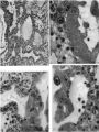
Plate 4
Plate 5
Plate 6
Plate 7
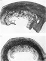
Plate 8
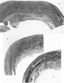
Plate 9
Plate 10
Plate 11
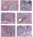
Plate 12
Plate 13


Reference
Hamilton WJ. and Boyd JD. Development of the human placenta in the first three months of gestation. (1960) J Anat. 94(3): 297-328. PMID14399291 | PDF
Cite this page: Hill, M.A. (2024, April 25) Embryology HamiltonBoyd1960 plate07.jpg. Retrieved from https://embryology.med.unsw.edu.au/embryology/index.php/File:HamiltonBoyd1960_plate07.jpg
- © Dr Mark Hill 2024, UNSW Embryology ISBN: 978 0 7334 2609 4 - UNSW CRICOS Provider Code No. 00098G
File history
Click on a date/time to view the file as it appeared at that time.
| Date/Time | Thumbnail | Dimensions | User | Comment | |
|---|---|---|---|---|---|
| current | 12:41, 6 August 2020 | | 1,280 × 1,536 (608 KB) | Z8600021 (talk | contribs) | crop, adjust size |
| 12:37, 6 August 2020 |  | 1,030 × 1,429 (450 KB) | Z8600021 (talk | contribs) |
You cannot overwrite this file.
File usage
The following 12 pages use this file:
- Paper - Development of the human placenta in the first three months of gestation (1960)
- File:HamiltonBoyd1960 fig02.jpg
- File:HamiltonBoyd1960 fig03.jpg
- File:HamiltonBoyd1960 fig04.jpg
- File:HamiltonBoyd1960 fig05.jpg
- File:HamiltonBoyd1960 fig06.jpg
- File:HamiltonBoyd1960 fig07.jpg
- File:HamiltonBoyd1960 fig08.jpg
- File:HamiltonBoyd1960 plate02.jpg
- File:HamiltonBoyd1960 plate03.jpg
- File:HamiltonBoyd1960 plate13.jpg
- Template:HamiltonBoyd1960 plates