File:HamiltonBoyd1960 fig02.jpg
Original file (800 × 836 pixels, file size: 121 KB, MIME type: image/jpeg)
Summary
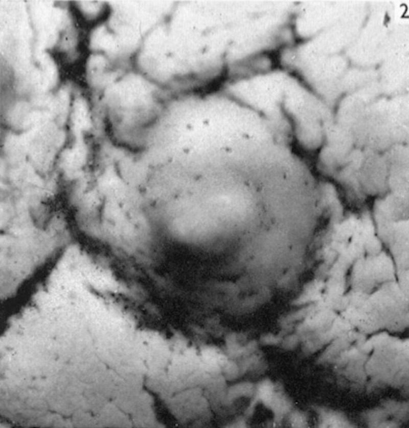
Fig. 2. Barnes Embryo
Photograph ( x 12) of the surface view of the implantation site of the Barnes embryo.
The implantation site itself appears as a slightly elevated area of the endometrium on which openings of the uterine glands can be seen. The surface of the endometrium shows characteristic shallow and irregular furrows.
Plates: 1 | 2 | 3 | 4 | 5 | 6 | 7 | 8 | 9 | 10 | 11 | 12 | 13
Plate 1
Plate 2
Plate 3
Plate 4
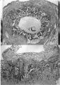
Plate 5
Plate 6
Plate 7
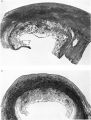
Plate 8
Plate 9
Plate 10
Plate 11
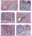
Plate 12
Plate 13


Reference
Hamilton WJ. and Boyd JD. Development of the human placenta in the first three months of gestation. (1960) J Anat. 94(3): 297-328. PMID14399291 | PDF
Cite this page: Hill, M.A. (2024, April 25) Embryology HamiltonBoyd1960 fig02.jpg. Retrieved from https://embryology.med.unsw.edu.au/embryology/index.php/File:HamiltonBoyd1960_fig02.jpg
- © Dr Mark Hill 2024, UNSW Embryology ISBN: 978 0 7334 2609 4 - UNSW CRICOS Provider Code No. 00098G
File history
Click on a date/time to view the file as it appeared at that time.
| Date/Time | Thumbnail | Dimensions | User | Comment | |
|---|---|---|---|---|---|
| current | 13:11, 6 August 2020 | | 800 × 836 (121 KB) | Z8600021 (talk | contribs) | ==Fig. 2. Barnes Embryo== Photograph ( x 12) of the surface view of the implantation site of the Barnes embryo. The implantation site itself appears as a slightly elevated area of the endometrium on which openings of the uterine glands can be seen. The surface of the endometrium shows characteristic shallow and irregular furrows. |
You cannot overwrite this file.
File usage
The following 2 pages use this file: