File:Hamilton1942 plate01.jpg

Original file (1,715 × 2,541 pixels, file size: 590 KB, MIME type: image/jpeg)
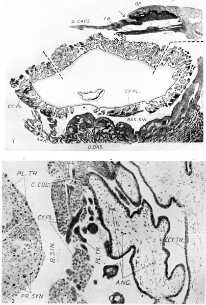
Plate 1
1. General view showing the relations of the embryonic rudiment and chorionic vesicle. The decidua basalis and a detached fragment of the decidua capsularis are also seen. The latter is traversed by the occlusion plug, and a part of the operculum is seen. The photograph was obtained from two original photographic prints which were pieced together and rephotographed so as to obtain a complete picture. An arrow indicates what we believe to be the point of entry. The interrupted line indicates the line of junction between the separate photographs. x c. 15.
2. Section through the wall of the chorionic vesicle and a part of the decidua basalis; showing the general relations of the villi, cytotrophoblastic plates and basal sinus. x c. 140.
Reference
Hamilton WJ. and Gladstone RJ. A presomite human embryo (Shaw) - the implantation. (1942) J Anat. 76(2): 187-203 PMID 17104888
Cite this page: Hill, M.A. (2024, April 24) Embryology Hamilton1942 plate01.jpg. Retrieved from https://embryology.med.unsw.edu.au/embryology/index.php/File:Hamilton1942_plate01.jpg
- © Dr Mark Hill 2024, UNSW Embryology ISBN: 978 0 7334 2609 4 - UNSW CRICOS Provider Code No. 00098G
File history
Click on a date/time to view the file as it appeared at that time.
| Date/Time | Thumbnail | Dimensions | User | Comment | |
|---|---|---|---|---|---|
| current | 16:07, 26 February 2017 | | 1,715 × 2,541 (590 KB) | Z8600021 (talk | contribs) | |
| 16:06, 26 February 2017 |  | 1,751 × 2,713 (1.36 MB) | Z8600021 (talk | contribs) | Hamilton1942 |
You cannot overwrite this file.
File usage
The following 2 pages use this file: