File:Haines1947 plate03.jpg

Original file (1,280 × 1,606 pixels, file size: 483 KB, MIME type: image/jpeg)
Plate 3
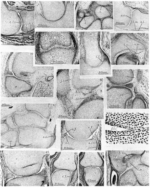
32. 30 mm. Appleton’s ‘H 30’, 5.8. Humero-radial joint. The middle layer of the interzone is liquefying and a small joint cavity has appeared anteriorly. The chondrogenic layers of the interzone ('c.l.) are still recognizable. The surface of the synovial mesenchyme is ragged. Loose tissue (l.t.) separates the annular ligament (a.l.) from the head of the radius.
33. 30 mm. Appleton’s ‘H 30’, 10.3. Humero-ulnar joint. This section is from the same series as that shown in fig. 32, and probably had a three-layered interzone, but as a result of pressure the interzone appears as a single dense layer of cells.
34. 30 mm. Appleton’s ‘H 30’. Carpal region. Most of the interzones are three layered in structure. The homogeneous appearance of some of the interzones (h.) is probably due to artifact.
35. West’s 34 mm. embryo, 12.2.4. Humero-radial joint. The humerus and radius are Between the head of the radius and the annular ligament (a.l.) is a layer of loose mesenchyme (l.m.) which will later break down to form a part of the joint cavity. 36. West’s 34 mm. embryo, 10.1.4. Humero-ulnar joint. Near the coronoid process the interzone has a typical three-layered structure, with a well-developed intermediate layer (i.l.). More posteriorly the intermediate layer appears as if compressed, an appearance probably due to artifact. . ' 37. West’s 34 mm. embryo, 10.1.3. Proximal inter-phalangeal joint of the fourth digit of the hand. The interzone is in a typical three-layered stage, with distinct chondrogenic layers (c.l.), and a loose intermediate layer (i.l.). This stage of the interzone has not usually been recognized in the inter-phalangeal joint.
38. West’s 34 mm. embryo, 20.1.5. Hip-joint. The head of the femur is seen in acetabulum, The ligamentum teres (l.t.) lies in a mass of synovial mesenchyme (s.m.) accompanied by conspicuous blood vessels (b.v.).
39. West’s 34 mm. embryo, 9.2.4. Knee. A small joint cavity has developed between the meniscus and the condyle of the femur, but the meniscus is still attached to the tibia by synovial mesenchyme.
40. West’s 34 mm. embryo, 6.2.5. Knee. A joint cavity (j.c.) is forming between the patella and femur. The anterior cruciate ligament (cr.) lies in a mass of synovial mesenchyme well supplied with blood vessels (b.v.). _
41. West’s 34 mm. embryo, 4.1.6. Knee. The tendon of the popliteus (pp) is seen in its characteristic position above the head of the fibula and outside the meniscus. The sesamoid in the lateral head of the gastrocnemius (am) is in the pro-cartilaginous stage. A small joint cavity (j.c.) is liquefying between the sesamoid and the femoral condyle.
42. West’s 34 mm. embryo, 9.1.4. Ankle. The interzones of the ankle and intertarsal joints are all in the three-layered stage. The synovial sheaths (s.s.) of the peroneus longus and Achilles’ tendon are already developed.
43. 45 mm. Appleton’s ‘H 45’, 4.2. Humero-radial joint. The humerus and radius appear joined, but this is probably an artifact. The joint cavity (j.c.) between the annular ligament (a.l.) and the head of the radius is well developed. Synovial folds (8. f.) project into the joint» and are covered by smooth strata of synovial cells (s.c.).
44. 45 mm. Appleton’s ‘H 45’, 4.2. Humero-radial joint. High-power view of the articular surfaces from the section illustrated in fig. 43. The surfaces are covered by fibrillar layers (f.l.), which unite where the surfaces join. Flat cells (f.c.) lie in and near these layers, and some cellular debris (d.) can be detected.
45. 45 mm. Appleton’s ‘H 45’, 26.1. Knee-joint. The menisci are free from the femur but still joined to the tibia by loose synovial mesenchyme.
46. 45 mm. Appleton’s ‘H 45’, 4.2. Metacarpo-phalangeal joint of the third digit of the hand. The cartilages appear joined. The collateral ligaments (c.l.) are seen on either side.
47. 45 mm. Appleton’s ‘H 45 ’, 10.6. Metacarpo-phalangeal joint of the fifth digit of the hand. In this section from the same series as fig. 46, the cartilages appear separate.
48. 49 mm. Appleton’s ‘H 49’, 33.1. Wrist. The fibro-cartilage (f.c.) of the wrist is found as a condensation of the synovial mesenchyme, with blood vessels (b.v.) nearby. A cartilage, the ‘intermedium antibrachii’ (i.a.), is found at this stage of development separated, from the styloid process of the ulna by a typical homogeneous interzone (h.z'.).
Reference
Haines RW. The development of joints. (1947) J. Anat. 81, 33-55.
Cite this page: Hill, M.A. (2024, April 20) Embryology Haines1947 plate03.jpg. Retrieved from https://embryology.med.unsw.edu.au/embryology/index.php/File:Haines1947_plate03.jpg
- © Dr Mark Hill 2024, UNSW Embryology ISBN: 978 0 7334 2609 4 - UNSW CRICOS Provider Code No. 00098G
File history
Click on a date/time to view the file as it appeared at that time.
| Date/Time | Thumbnail | Dimensions | User | Comment | |
|---|---|---|---|---|---|
| current | 15:04, 3 October 2017 |  | 1,280 × 1,606 (483 KB) | Z8600021 (talk | contribs) | |
| 15:03, 3 October 2017 |  | 1,748 × 2,413 (783 KB) | Z8600021 (talk | contribs) |
You cannot overwrite this file.
File usage
The following page uses this file: