File:Haines1947 plate01.jpg

Original file (1,280 × 1,653 pixels, file size: 544 KB, MIME type: image/jpeg)
Plate 1
fig. 1. 10 mm. Lucas Keene’s ‘2387’, 50.4. Fore-limb. The skeletal blastema (sIc.) shows clearly in the humeral, elbow (el.) and fore-arm regions, while distally it fades away towards the marginal vein. The pre-muscle masses of the upper arm on both flexor (fl.) and extensor (e:c.) aspects, the brachial plexus ( pl.) and median nerve (md.) are visible. A blood vessel of the interosseous group (b.v.) pierces the blastema.
fig. 2. [10-5 mm. Smout’s 5.5 weeks embryo, 11.1.4. Fore-limb. The pre-muscle masses of the lower arm have now differentiated, and the boundaries of the skeletal blastema are less easy to follow, as the condensations for the skeleton and the muscles now form a continuous mass.
fig. 3. 10-5 mm. Smout’s 5.5 weeks embryo, 16.1.4. Hind-limb. The skeletal blastema of the femoral (fm.), tibial (tb.) and foot-plate (f.p.) regions are distinguishable. The pre-muscle masses of the thigh (p.m.) are more condensed than the general mesenchyme, but those for the more distal part of the limbs have not yet appeared.
fig. 4. 12 mm. Smout’s 6 weeks embryo, 31.3.5. Humero-radial joint. The lower end of the cartilaginous humerus (hm.) is assuming its characteristic shape and the shaft of the radius (rd.) is well developed, but the ends are not yet chondrified. A thick interzone (i.z.) separates the two chondrifications and gives the first indication of joint formation. Peripherally the interzone is continuous with the dense mesenchyme surrounding the cartilages which now forms their perichondria (p.c.). Distally the blastema of the radius is quite continuous with that of the carpal region and with the condensed tissue which will form the muscular and tendinous apparatus. ,
5. 12 mm. Smout’s 6 weeks embryo, 49.2.5. Thigh. The shaft of the femur (fm.) is well developed, but that of the tibia (tb.) is barely recognizable as a small centre of chondrification. The knee appears as a thick interzone (i.z.) but at the hip the femoral tissues are continued without interruption into the dense blastema of the pelvis (pl.).
6. 12 mm. Smout’s 6 weeks embryo, 33.1.5. Carpal region. The shaft of the ulna (ul.) is well formed and distinct from the muscles on each side, and the carpal region is formed by a dense mass of blastema in which the carpal elements are appearing as regions of greater density (cp.). The metacarpal region shows early centres of chondrification (mt.), but passes without any break into the blastema for the phalangeal region (ph.) and the dense tissues destined to form the flexor and extensor apparatus.
7. 13 mm. Boyd’s ‘H 23’, 24.1.5. Shoulder joint. The scapula with its characteristic blade (bl.) and coracoid (cr.) is well chondrified, and is separated from the head of the humerus (hm.) by an interzone»(i.z.). The interzone is continuous at its margins with the perichondrium (p.c.) of the scapula and humerus, and through this with the very dense tissues surrounding the head of the humerus in which the tendons of the short muscles of the joint and the long head of the biceps will differentiate. The muscular belly of the deltoid (dl.) and its nerve supply (n.s.) are quite distinct. ,
8. 13 mm. Boyd’s ‘H 23’, 23.3.4. Humero-ulnar joint. The trochlear region of the humerus (hm.) is separated from the ulnar (ul.) by a thick interzone (z'.z.). The ulna has not -yet developed its olecranon and coronoid processes, though the triceps (tr.) is quite distinct, and there is little indication of the future shape of the joint surfaces.
9. 13 mm. Boyd’s ‘H 23’, 24.1.6. Carpus. The lower end of the radius (r.d.) is separated from the metacarpal region (mt.) by two masses of condensed blastemal tissue, which represent the proximal and distal rows of carpal elements (cp.). Between these masses the tissue is rarefied, and not condensed as yet to form typical interzones.
10. 13 mm. Boyd’s ‘H 23’, 40.2.4. Lower limb. The femur is now well defined and the lower end fully chondrified. The hip bone is chondrified and separated from the femur by an interzone. The tibia is well developed and the interzone at the knee has become sharply defined. The quadriceps muscle mass (qu.) surrounds the femoral nerve (f.n.), and the quadriceps tendon (q.t.) is seen passing in front of the knee joint region, quite distinct from it. The hamstring mass (hm.) surrounds the sciatic nerve (s.n.) in a similar way, and is also distinct from the skeletal tissues, being separated by a layer of connective tissue (c.t.). In the foot chondrification has not yet begun. In the tarsal region the elements are represented by dense masses of blastemal tissue (ts.). The metatarsal and phalangeal regions of the blastema (mt. and ph.) are not yet differentiated.
11. 14 mm. Boyd’s ‘H 8’, 19.2.8. Humero-ulnar joint. The trochlear surface of the humerus is separated from the sigmoid notch of the ulna by a well-defined interzone (i.z.) which follows the shape of the articular surfaces. The olecranon (ol.) and coronoid (cr.) processes of the ulna are now distinct. The interzone, perichondrium and tissues concerned with the tendons near the joint are still intimately blended.
12. 14 mm. Boyd’s ‘H 8’, 19.3.8. Fore-arm and hand. The carpallelements of the proximal and distal rows (cp.) are now chondrified, and are separated from each other and from the radius (rd.) and metacarpals (mt.) by typical interzones. The phalangeal region of the blastema (ph.) is still undifferentiated.
13. 16 mm. Kirk’s ‘Series 1 ’, 22.2.6. Shoulder. The interzone passes peripherally into the dense tissue (d.t.) that will form the capsule, labrum and neighbouring tendons, but these structures are not yet differentiated. Small blood vessels (b.v.) lie outside the perichondrium, forming a network which follows the shape of the cartilages, but none enters the perichondrium itself.
14. 16 mm. Kirk’s ‘Series 1 ’, 23.4.3. Humero-radial joint. Anteriorly and posteriorly the fibrous capsule (f.c.) arches over the joint region, enclosing the synovial mesachyme (s.m.) between its inner surface and the intra-capsular perichondrium (5.1).). Small blood vessels lie within the capsule.
Reference
Haines RW. The development of joints. (1947) J. Anat. 81, 33-55.
Cite this page: Hill, M.A. (2024, April 25) Embryology Haines1947 plate01.jpg. Retrieved from https://embryology.med.unsw.edu.au/embryology/index.php/File:Haines1947_plate01.jpg
- © Dr Mark Hill 2024, UNSW Embryology ISBN: 978 0 7334 2609 4 - UNSW CRICOS Provider Code No. 00098G
File history
Click on a date/time to view the file as it appeared at that time.
| Date/Time | Thumbnail | Dimensions | User | Comment | |
|---|---|---|---|---|---|
| current | 15:14, 3 October 2017 | | 1,280 × 1,653 (544 KB) | Z8600021 (talk | contribs) | |
| 15:13, 3 October 2017 | 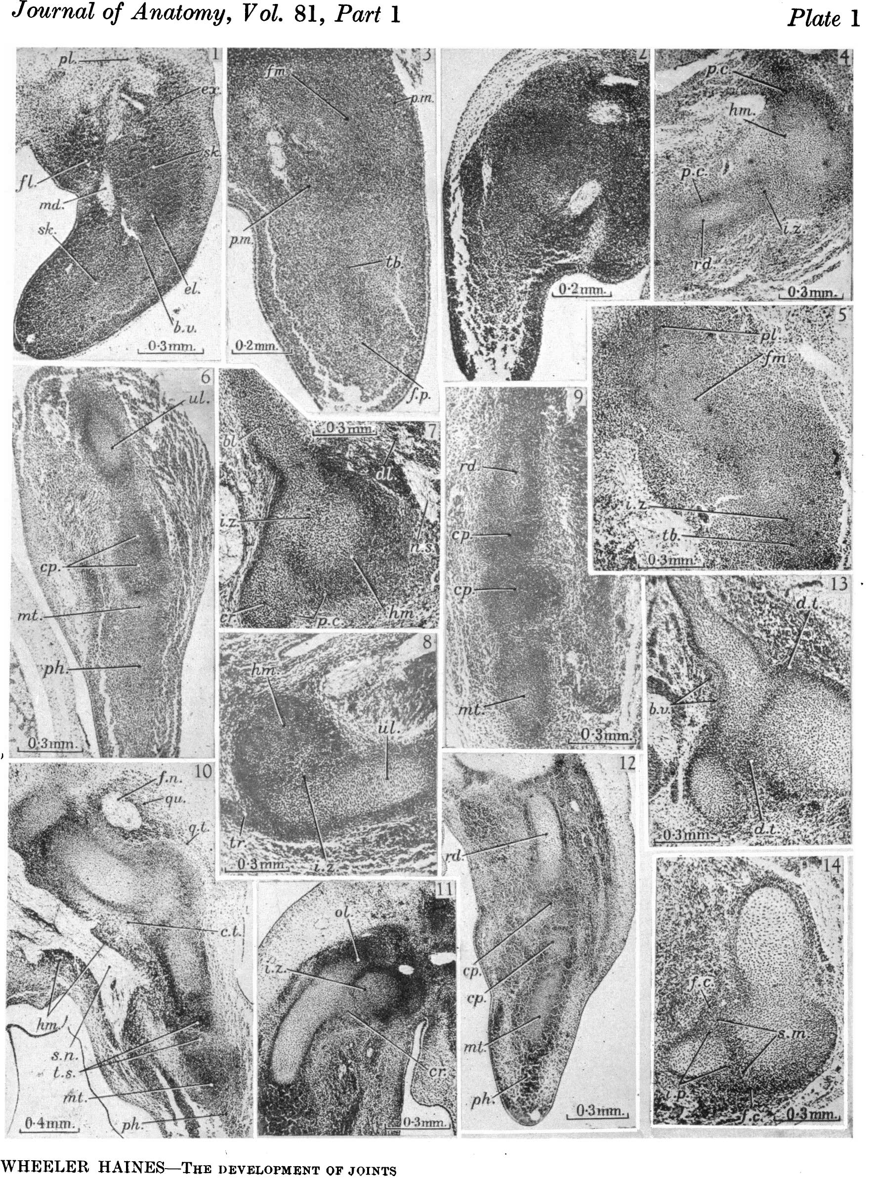 | 1,767 × 2,391 (880 KB) | Z8600021 (talk | contribs) | ==Plate 1== |
You cannot overwrite this file.
File usage
The following page uses this file: