File:Gray1181.jpg
Gray1181.jpg (800 × 487 pixels, file size: 77 KB, MIME type: image/jpeg)
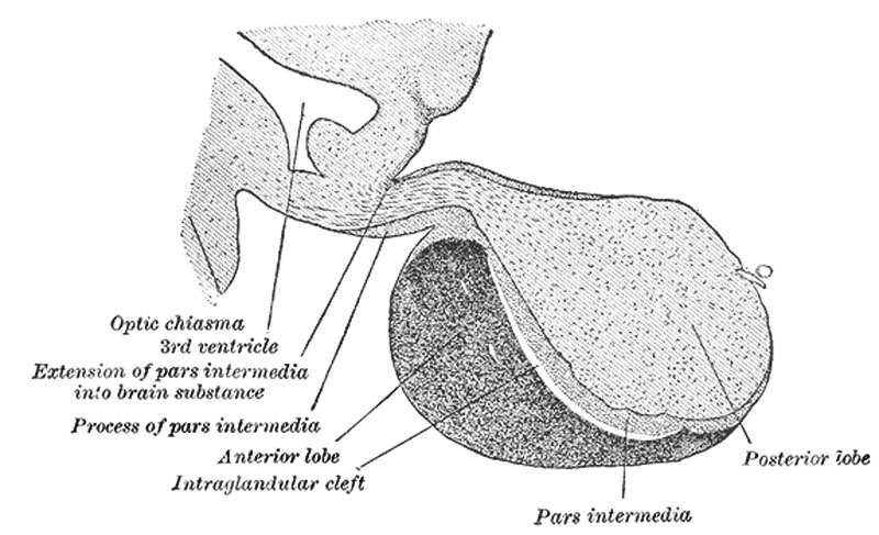
Fig. 1181. Pituitary - Historic drawing of median sagittal through the hypophysis of an adult monkey
Median sagittal through the hypophysis of an adult monkey. Semidiagrammatic. (Herring.)
The hypophysis consists of an anterior and a posterior lobe, which differ from one another in their mode of development and in their structure (Fig. 1181).
The anterior lobe is the larger and is somewhat kidney-shaped, the concavity being directed backward and embracing the posterior lobe. It consists of a pars anterior and a pars intermedia, separated from each other by a narrow cleft, the remnant of the pouch or diverticulum. The pars anterior is extremely vascular and consists of epithelial cells of varying size and shape, arranged in cord-like trabeculæ or alveoli and separated by large, thin-walled blood vessels.
The pars intermedia is a thin lamina closely applied to the body and neck of the posterior lobe and extending onto the neighboring parts of the brain; it contains few bloodvessels and consists of finely granular cells between which are small masses of colloid material.
The pars intermedia in spite of the fact that it arises in common with the pars anterior from the ectoderm of the primitive buccal cavity is often (incorrectly) considered as a part of the posterior lobe which arises from the floor of the third ventricle of the brain.
(Modified Gray's text)
- median eminence
- infundibulum - short attaching stalk.
- pars tuberalis - wrapped around infundibulum.
- Links: Pituitary Development | Medicine Endocrine Lecture | Historic pituitary drawing | Image - hypophysis of an adult monkey
- Gray's Images: Development | Lymphatic | Neural | Vision | Hearing | Somatosensory | Integumentary | Respiratory | Gastrointestinal | Urogenital | Endocrine | Surface Anatomy | iBook | Historic Disclaimer
| Historic Disclaimer - information about historic embryology pages |
|---|
| Pages where the terms "Historic" (textbooks, papers, people, recommendations) appear on this site, and sections within pages where this disclaimer appears, indicate that the content and scientific understanding are specific to the time of publication. This means that while some scientific descriptions are still accurate, the terminology and interpretation of the developmental mechanisms reflect the understanding at the time of original publication and those of the preceding periods, these terms, interpretations and recommendations may not reflect our current scientific understanding. (More? Embryology History | Historic Embryology Papers) |
| iBook - Gray's Embryology | |
|---|---|

|
|
Reference
Gray H. Anatomy of the human body. (1918) Philadelphia: Lea & Febiger.
Cite this page: Hill, M.A. (2024, April 19) Embryology Gray1181.jpg. Retrieved from https://embryology.med.unsw.edu.au/embryology/index.php/File:Gray1181.jpg
- © Dr Mark Hill 2024, UNSW Embryology ISBN: 978 0 7334 2609 4 - UNSW CRICOS Provider Code No. 00098G
File history
Click on a date/time to view the file as it appeared at that time.
| Date/Time | Thumbnail | Dimensions | User | Comment | |
|---|---|---|---|---|---|
| current | 16:09, 15 May 2012 |  | 800 × 487 (77 KB) | Z8600021 (talk | contribs) | |
| 17:38, 9 June 2011 |  | 600 × 376 (53 KB) | MarkHill (talk | contribs) | ==Pituitary - Median sagittal through the hypophysis of an adult monkey== Median sagittal through the hypophysis of an adult monkey. Semidiagrammatic. (Herring.) {{Gray Anatomy}} |
You cannot overwrite this file.
File usage
The following 3 pages use this file:

