File:Gray1175.jpg
Gray1175.jpg (500 × 500 pixels, file size: 31 KB, MIME type: image/jpeg)
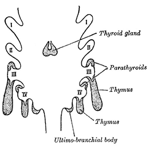
Scheme showing development of branchial epithelial bodies
(Modified from Koh.) I, II, III, IV. Branchial pouches.
Development
The thyroid gland is developed from a median diverticulum (Fig. 1175), which appears about the fourth week on the summit of the tuberculum impar, but later is found in the furrow immediately behind the tuberculum (Fig. 979).
It grows downward and backward as a tubular duct, which bifurcates and subsequently subdivides into a series of cellular cords, from which the isthmus and lateral lobes of the thyroid gland are developed. The ultimo-branchial bodies from the fifth pharyngeal pouches are enveloped by the lateral lobes of the thyroid gland; they undergo atrophy and do not form true thyroid tissue.
The connection of the diverticulum with the pharynx is termed the thyroglossal duct; its continuity is subsequently interrupted, and it undergoes degeneration, its upper end being represented by the foramen cecum of the tongue, and its lower by the pyramidal lobe of the thyroid gland.
Online Editor - thymus note in contrast to this historic image, current research has shown that the human thymic epithelium derives solely from the third pharyngeal pouch (as in the mouse).
Farley AM, Morris LX, Vroegindeweij E, Depreter ML, Vaidya H, Stenhouse FH, Tomlinson SR, Anderson RA, Cupedo T, Cornelissen JJ & Blackburn CC. (2013). Dynamics of thymus organogenesis and colonization in early human development. Development , 140, 2015-26. PMID: 23571219 DOI.
- Gray's Images: Development | Lymphatic | Neural | Vision | Hearing | Somatosensory | Integumentary | Respiratory | Gastrointestinal | Urogenital | Endocrine | Surface Anatomy | iBook | Historic Disclaimer
| Historic Disclaimer - information about historic embryology pages |
|---|
| Pages where the terms "Historic" (textbooks, papers, people, recommendations) appear on this site, and sections within pages where this disclaimer appears, indicate that the content and scientific understanding are specific to the time of publication. This means that while some scientific descriptions are still accurate, the terminology and interpretation of the developmental mechanisms reflect the understanding at the time of original publication and those of the preceding periods, these terms, interpretations and recommendations may not reflect our current scientific understanding. (More? Embryology History | Historic Embryology Papers) |
| iBook - Gray's Embryology | |
|---|---|

|
|
Reference
Gray H. Anatomy of the human body. (1918) Philadelphia: Lea & Febiger.
Cite this page: Hill, M.A. (2024, April 25) Embryology Gray1175.jpg. Retrieved from https://embryology.med.unsw.edu.au/embryology/index.php/File:Gray1175.jpg
- © Dr Mark Hill 2024, UNSW Embryology ISBN: 978 0 7334 2609 4 - UNSW CRICOS Provider Code No. 00098G
File history
Click on a date/time to view the file as it appeared at that time.
| Date/Time | Thumbnail | Dimensions | User | Comment | |
|---|---|---|---|---|---|
| current | 12:29, 25 February 2013 |  | 500 × 500 (31 KB) | Z8600021 (talk | contribs) | ==Scheme showing development of branchial epithelial bodies== (Modified from Koh.) I, II, III, IV. Branchial pouches. Development.—The thyroid gland is developed from a median diverticulum (Fig. 1175), which appears about the fourth week on the summit |
You cannot overwrite this file.
File usage
The following page uses this file:

