File:Gray1125.jpg
Gray1125.jpg (500 × 584 pixels, file size: 69 KB, MIME type: image/jpeg)
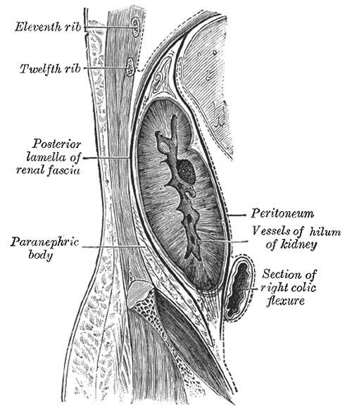
Sagittal section through posterior abdominal wall, showing the relations of the capsule of the kidney
After Gerota
Fixation of the Kidney (Figs. 1125, 1126).
The kidney and its vessels are imbedded in a mass of fatty tissue, termed the adipose capsule, which is thickest at the margins of the kidney and is prolonged through the hilum into the renal sinus. The kidney and the adipose capsule are enclosed in a sheath of fibrous tissue continuous with the subperitoneal fascia, and named the renal fascia. At the lateral border of the kidney the renal fascia splits into an anterior and a posterior layer. The anterior layer is carried medialward in front of the kidney and its vessels, and is continuous over the aorta with the corresponding layer of the opposite side. The posterior layer extends medialward behind the kidney and blends with the fascia on the Quadratus lumborum and Psoas major, and through this fascia is attached to the vertebral column. Above the suprarenal gland the two layers of the renal fascia fuse, and unite with the fascia of the diaphragm; below they remain separate, and are gradually lost in the subperitoneal fascia of the iliac fossa. The renal fascia is connected to the fibrous tunic of the kidney by numerous trabeculæ, which traverse the adipose capsule, and are strongest near the lower end of the organ. Behind the fascia renalis is a considerable quantity of fat, which constitutes the paranephric body. The kidney is held in position partly through the attachment of the renal fascia and partly by the apposition of the neighboring viscera.
- Links: Renal System Development
- Gray's Images: Development | Lymphatic | Neural | Vision | Hearing | Somatosensory | Integumentary | Respiratory | Gastrointestinal | Urogenital | Endocrine | Surface Anatomy | iBook | Historic Disclaimer
| Historic Disclaimer - information about historic embryology pages |
|---|
 |
| iBook - Gray's Embryology | |
|---|---|

|
|
Reference
Gray H. Anatomy of the human body. (1918) Philadelphia: Lea & Febiger.
Cite this page: Hill, M.A. (2024, April 18) Embryology Gray1125.jpg. Retrieved from https://embryology.med.unsw.edu.au/embryology/index.php/File:Gray1125.jpg
- © Dr Mark Hill 2024, UNSW Embryology ISBN: 978 0 7334 2609 4 - UNSW CRICOS Provider Code No. 00098G
File history
Click on a date/time to view the file as it appeared at that time.
| Date/Time | Thumbnail | Dimensions | User | Comment | |
|---|---|---|---|---|---|
| current | 23:02, 17 September 2012 | | 500 × 584 (69 KB) | Z8600021 (talk | contribs) | :'''Links:''' Renal System Development {{Template:Gray Anatomy}} Category:Renal |
You cannot overwrite this file.
File usage
The following page uses this file: