File:Gray1118.jpg
From Embryology
Gray1118.jpg (600 × 403 pixels, file size: 45 KB, MIME type: image/jpeg)
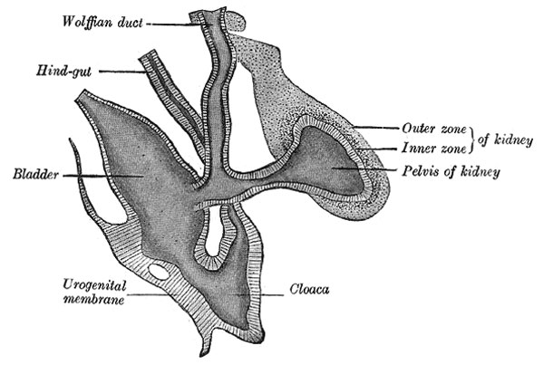
Primitive Kidney and Bladder
From a reconstruction (After Schreiner.)
The Urinary Bladder
- bladder is formed partly from the entodermal cloaca and partly from the ends of the Wolffian (mesonephric) ducts
- allantois takes no share in its formation
- after the separation of the rectum from the dorsal part of the cloaca the ventral part becomes subdivided into three portions
- an anterior vesico-urethral portion, continuous with the allantois - into this portion the Wolffian ducts open
- an intermediate narrow channel, the pelvic portion
- a posterior phallic portion, closed externally by the urogenital membrane
- the second and third parts together constitute the urogenital sinus
- the vesico-urethral portion absorbs the ends of the Wolffian (mesonephric) ducts and the associated ends of the renal diverticula
- these give rise to the trigone of the bladder and part of the prostatic urethra
- the remainder of the vesico-urethral portion forms the body of the bladder and part of the prostatic urethra
- its apex is prolonged to the umbilicus as a narrow canal
- later is obliterated and becomes the medial umbilical ligament (urachus)
(text modified from Gray's Anatomy)
- Gray's Images: Development | Lymphatic | Neural | Vision | Hearing | Somatosensory | Integumentary | Respiratory | Gastrointestinal | Urogenital | Endocrine | Surface Anatomy | iBook | Historic Disclaimer
| Historic Disclaimer - information about historic embryology pages |
|---|
| Pages where the terms "Historic" (textbooks, papers, people, recommendations) appear on this site, and sections within pages where this disclaimer appears, indicate that the content and scientific understanding are specific to the time of publication. This means that while some scientific descriptions are still accurate, the terminology and interpretation of the developmental mechanisms reflect the understanding at the time of original publication and those of the preceding periods, these terms, interpretations and recommendations may not reflect our current scientific understanding. (More? Embryology History | Historic Embryology Papers) |
| iBook - Gray's Embryology | |
|---|---|

|
|
Reference
Gray H. Anatomy of the human body. (1918) Philadelphia: Lea & Febiger.
Cite this page: Hill, M.A. (2024, April 25) Embryology Gray1118.jpg. Retrieved from https://embryology.med.unsw.edu.au/embryology/index.php/File:Gray1118.jpg
- © Dr Mark Hill 2024, UNSW Embryology ISBN: 978 0 7334 2609 4 - UNSW CRICOS Provider Code No. 00098G
File history
Click on a date/time to view the file as it appeared at that time.
| Date/Time | Thumbnail | Dimensions | User | Comment | |
|---|---|---|---|---|---|
| current | 08:21, 28 May 2011 |  | 600 × 403 (45 KB) | S8600021 (talk | contribs) | ==Primitive Kidney and Bladder== from a reconstruction (After Schreiner.) {{Historic Disclaimer}} ===The Urinary Bladder=== * bladder is formed partly from the entodermal cloaca and partly from the ends of the Wolffian ducts * allantois takes no share |
You cannot overwrite this file.
File usage
The following 5 pages use this file:

