File:Gray1110.jpg
Original file (414 × 1,200 pixels, file size: 86 KB, MIME type: image/jpeg)
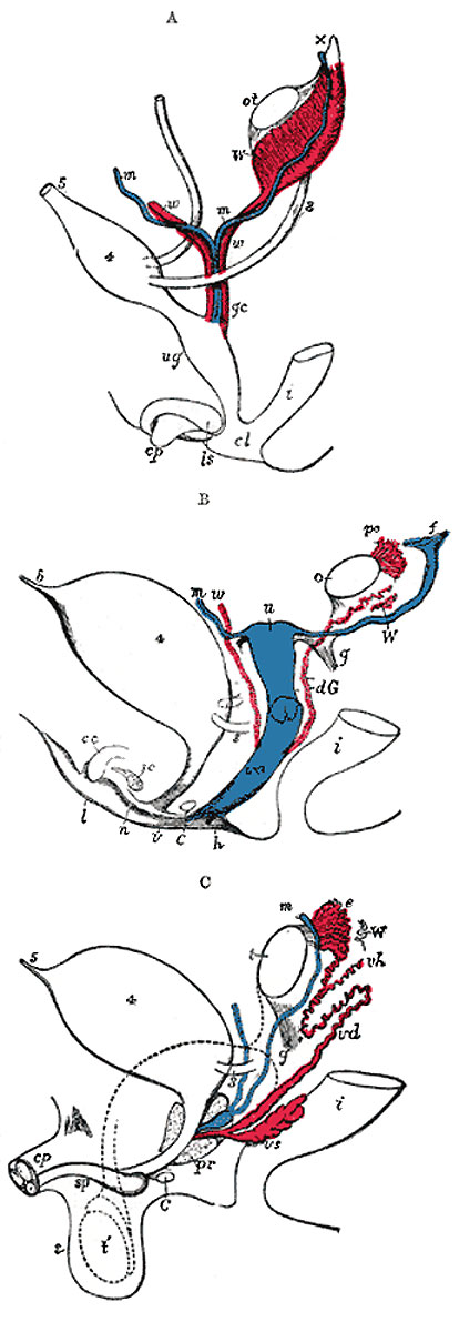
Diagrams to show the development of male and female generative organs from a common type
(Allen Thomson.)
Figure 1110: Fig. 1110a Indifferent | Fig. 1110b Female | Fig. 1110c Male
A Diagram of the primitive urogenital organs in the embryo previous to sexual distinction
3. Ureter. 4. Urinary bladder. 5. Urachus. cl. Cloaca. cp. Elevation which becomes clitoris or penis. i. Lower part of the intestine. ls. Fold of integument from which the labia majora or scrotum are formed. m, m. Right and left Müllerian ducts uniting together and running with the Wolffian ducts in gc, the genital cord. ot. The genital ridge from which either the ovary or testis is formed. ug. Sinus urogenitalis. W. Left Wolffian body. w, w. Right and left Wolffian ducts.
B Diagram of the female type of sexual organs
C. Greater vestibular gland, and immediately above it the urethra. cc. Corpus cavernosum clitoridis. dG. Remains of the left Wolffian duct, such as give rise to the duct of Gärtner, represented by dotted lines; that of the right side is marked w. f. The abdominal opening of the left uterine tube. g. Round ligament, corresponding to gubernaculum. h. Situation of the hymen. i. Lower part of the intestine. l. Labium major. n. Labium minus. o. The left ovary. po. Epoophoron. sc. Corpus cavernosum urethrae. u. Uterus. The uterine tube of the right side is marked m. v. Vulva. va. Vagina. W. Scattered remains of Wolffian tubes near it (paroöphoron of Waldeyer).
C Diagram of the male type of sexual organs
C. Bulbo-urethral gland of one side. cp. Corpora cavernosa penis cut short. e. Caput epididymis. g. The gubernaculum. i. Lower part of the intestine. m. Müllerian duct, the upper part of which remains as the hydatid of Morgagni; the lower part, represented by a dotted line descending to the prostatic utricle, constitutes the occasionally existing cornu and tube of the uterus masculinus. pr. The prostate. s. Scrotum. sp. Corpus cavernosum urethrae. t. Testis in the place of its original formation. t’, together with the dotted lines above, indicates the direction in which the testis and epididymis descend from the abdomen into the scrotum. vd. Ductus deferens. vh. Ductus aberrans. vs. The vesicula seminalis. W. Scattered remains of the Wolffian body, constituting the organ of Giraldès, or the paradidymis of Waldeyer.
- Genital Image Links: Fig. 1110 | Fig. 1110a Indifferent | Fig. 1110b Female | Fig. 1110c Male | Indifferent unlabeled | Female unlabeled | Male unlabeled | Genital System Development
- Gray's Images: Development | Lymphatic | Neural | Vision | Hearing | Somatosensory | Integumentary | Respiratory | Gastrointestinal | Urogenital | Endocrine | Surface Anatomy | iBook | Historic Disclaimer
| Historic Disclaimer - information about historic embryology pages |
|---|
 |
| iBook - Gray's Embryology | |
|---|---|

|
|
Reference
Gray H. Anatomy of the human body. (1918) Philadelphia: Lea & Febiger.
Cite this page: Hill, M.A. (2024, April 25) Embryology Gray1110.jpg. Retrieved from https://embryology.med.unsw.edu.au/embryology/index.php/File:Gray1110.jpg
- © Dr Mark Hill 2024, UNSW Embryology ISBN: 978 0 7334 2609 4 - UNSW CRICOS Provider Code No. 00098G
Cite this page: Hill, M.A. (2024, April 25) Embryology Gray1110.jpg. Retrieved from https://embryology.med.unsw.edu.au/embryology/index.php/File:Gray1110.jpg
- © Dr Mark Hill 2024, UNSW Embryology ISBN: 978 0 7334 2609 4 - UNSW CRICOS Provider Code No. 00098G
File history
Click on a date/time to view the file as it appeared at that time.
| Date/Time | Thumbnail | Dimensions | User | Comment | |
|---|---|---|---|---|---|
| current | 16:41, 4 June 2013 | 414 × 1,200 (86 KB) | Z8600021 (talk | contribs) | ||
| 16:35, 4 June 2013 | 414 × 1,200 (75 KB) | Z8600021 (talk | contribs) |
You cannot overwrite this file.
File usage
There are no pages that use this file.