File:Gray1050.jpg
Gray1050.jpg (765 × 500 pixels, file size: 92 KB, MIME type: image/jpeg)
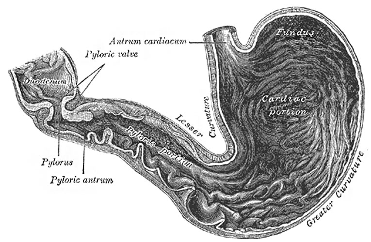
Fig. 1050. Interior of the Stomach
When examined after death, the stomach is usually fixed at some temporary stage of the digestive process. A common form is that shown in Fig. 1050.
If the organ (viscus) be laid open by a section through the plane of its two curvatures, it is seen to consist of two segments: (a) a large globular portion on the left and (b) a narrow tubular part on the right. These correspond to the clinical subdivisions of fundus and pyloric portions already described, and are separated by a constriction which indents the body and greater curvature, but does not involve the lesser curvature. To the left of the cardiac orifice is the incisura cardiaca: the projection of this notch into the cavity of the stomach increases as the organ distends, and has been supposed to act as a valve preventing regurgitation into the esophagus.
In the pyloric portion are seen: (a) the elevation corresponding to the incisura angularis, and (b) the circular projection from the duodenopyloric constriction which forms the pyloric valve; the separation of the pyloric antrum from the rest of the pyloric part is scarcely indicated.
- Gray's Images: Development | Lymphatic | Neural | Vision | Hearing | Somatosensory | Integumentary | Respiratory | Gastrointestinal | Urogenital | Endocrine | Surface Anatomy | iBook | Historic Disclaimer
| Historic Disclaimer - information about historic embryology pages |
|---|
| Pages where the terms "Historic" (textbooks, papers, people, recommendations) appear on this site, and sections within pages where this disclaimer appears, indicate that the content and scientific understanding are specific to the time of publication. This means that while some scientific descriptions are still accurate, the terminology and interpretation of the developmental mechanisms reflect the understanding at the time of original publication and those of the preceding periods, these terms, interpretations and recommendations may not reflect our current scientific understanding. (More? Embryology History | Historic Embryology Papers) |
| iBook - Gray's Embryology | |
|---|---|

|
|
Reference
Gray H. Anatomy of the human body. (1918) Philadelphia: Lea & Febiger.
Cite this page: Hill, M.A. (2024, April 25) Embryology Gray1050.jpg. Retrieved from https://embryology.med.unsw.edu.au/embryology/index.php/File:Gray1050.jpg
- © Dr Mark Hill 2024, UNSW Embryology ISBN: 978 0 7334 2609 4 - UNSW CRICOS Provider Code No. 00098G
File history
Click on a date/time to view the file as it appeared at that time.
| Date/Time | Thumbnail | Dimensions | User | Comment | |
|---|---|---|---|---|---|
| current | 13:10, 11 May 2014 |  | 765 × 500 (92 KB) | Z8600021 (talk | contribs) | ==Fig. 1050. Interior of the Stomach== Category:Stomach |
You cannot overwrite this file.
File usage
The following 3 pages use this file:

