File:Gray1042.jpg

Original file (969 × 745 pixels, file size: 140 KB, MIME type: image/jpeg)
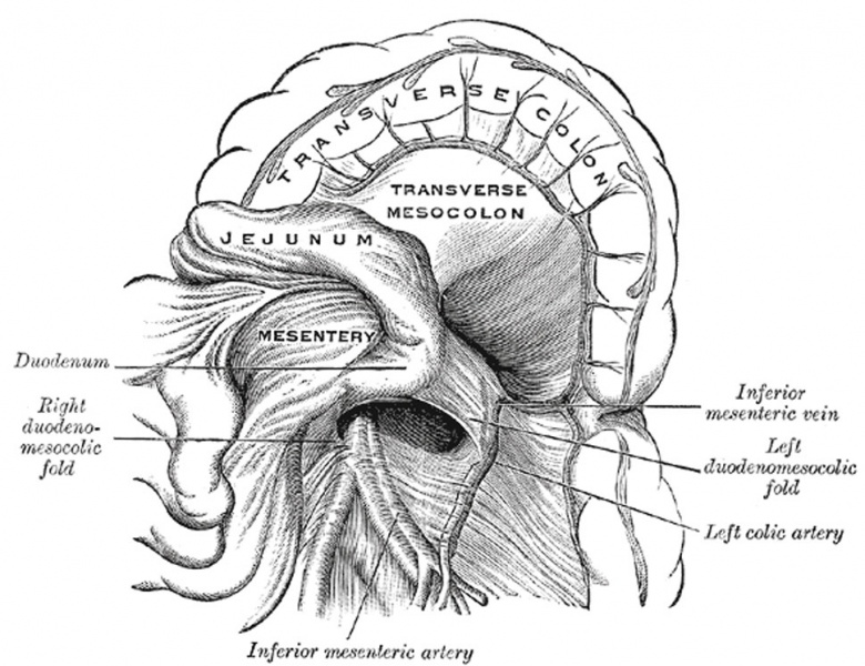
Duodenojejunal Fossa
(Poirier and Charpy.)
The duodenojejunal fossa exists in from 15 to 20 per cent. of cases, but has never yet been found in conjunction with the other forms of duodenal fossæ it can be seen by pulling the jejunum downward and to the right, after the transverse colon has been pulled upward. It is bounded above by the pancreas, to the right by the aorta, and to the left by the kidney; beneath is the left renal vein. It has a depth of from 2 to 3 cm., and its orifice, directed downward and to the right, is nearly circular and will admit the tip of the little finger.
Peritoneal Recesses or Fossæ
(retroperitoneal fossæ) In certain parts of the abdominal cavity there are recesses of peritoneum forming culs-de-sac or pouches, which are of surgical interest in connection with the possibility of the occurrence of “retroperitoneal” herniæ. The largest of these is the omental bursa (already described), but several others, of smaller size, require mention, and may be divided into three groups, viz.: duodenal, cecal, and intersigmoid.Duodenal Fossæ (Figs. 1041, 1042)
Three are fairly constant:
(a) The inferior duodenal fossa, present in from 70 to 75 per cent. of cases, is situated opposite the third lumbar vertebra on the left side of the ascending portion of the duodenum. Its opening is directed upward, and is bounded by a thin sharp fold of peritoneum with a concave margin, called the duodenomesocolic fold. The tip of the index finger introduced into the fossa under the fold passes some little distance behind the ascending portion of the duodenum.
(b) The superior duodenal fossa, present in from 40 to 50 per cent. of cases, often coexists with the inferior one, and its orifice looks downward. It lies on the left of the ascending portion of the duodenum, in front of the second lumbar vertebra, and behind a sickle-shaped fold of peritoneum, the duodenojejunal fold, and has a depth of about 2 cm.
(c) The duodenojejunal fossa exists in from 15 to 20 per cent. of cases, but has never yet been found in conjunction with the other forms of duodenal fossæ it can be seen by pulling the jejunum downward and to the right, after the transverse colon has been pulled upward. It is bounded above by the pancreas, to the right by the aorta, and to the left by the kidney; beneath is the left renal vein. It has a depth of from 2 to 3 cm., and its orifice, directed downward and to the right, is nearly circular and will admit the tip of the little finger.
- Gray's Images: Development | Lymphatic | Neural | Vision | Hearing | Somatosensory | Integumentary | Respiratory | Gastrointestinal | Urogenital | Endocrine | Surface Anatomy | iBook | Historic Disclaimer
| Historic Disclaimer - information about historic embryology pages |
|---|
| Pages where the terms "Historic" (textbooks, papers, people, recommendations) appear on this site, and sections within pages where this disclaimer appears, indicate that the content and scientific understanding are specific to the time of publication. This means that while some scientific descriptions are still accurate, the terminology and interpretation of the developmental mechanisms reflect the understanding at the time of original publication and those of the preceding periods, these terms, interpretations and recommendations may not reflect our current scientific understanding. (More? Embryology History | Historic Embryology Papers) |
| iBook - Gray's Embryology | |
|---|---|

|
|
Reference
Gray H. Anatomy of the human body. (1918) Philadelphia: Lea & Febiger.
Cite this page: Hill, M.A. (2024, April 25) Embryology Gray1042.jpg. Retrieved from https://embryology.med.unsw.edu.au/embryology/index.php/File:Gray1042.jpg
- © Dr Mark Hill 2024, UNSW Embryology ISBN: 978 0 7334 2609 4 - UNSW CRICOS Provider Code No. 00098G
File history
Click on a date/time to view the file as it appeared at that time.
| Date/Time | Thumbnail | Dimensions | User | Comment | |
|---|---|---|---|---|---|
| current | 17:13, 30 April 2011 |  | 969 × 745 (140 KB) | S8600021 (talk | contribs) | ==Duodenojejunal Fossa== (Poirier and Charpy.) The duodenojejunal fossa exists in from 15 to 20 per cent. of cases, but has never yet been found in conjunction with the other forms of duodenal fossæ it can be seen by pulling the jejunum downward and |
You cannot overwrite this file.
File usage
There are no pages that use this file.
