File:Gray1040.jpg

Original file (900 × 918 pixels, file size: 230 KB, MIME type: image/jpeg)
Peritoneum Leaving Abdominal Wall
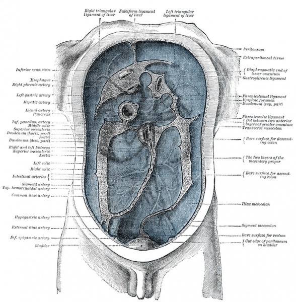
Diagram devised by Delépine to show the lines along which the peritoneum leaves the wall of the abdomen to invest the viscera.
The mesenteries are: the mesentery proper, the transverse mesocolon, and the sigmoid mesocolon. In addition to these there are sometimes present an ascending and a descending mesocolon.
mesentery proper
- mesentery proper (mesenterium) is the broad, fan-shaped fold of peritoneum
- connects the convolutions of the jejunum and ileum with the posterior wall of the abdomen
- root (part connected with the structures in front of the vertebral column) is narrow, about 15 cm. long, and is directed obliquely from the duodenojejunal flexure at the left side of the second lumbar vertebra to the right sacroiliac articulation (Fig. 1040)
- intestinal border is about 6 metres long
- here the two layers separate to enclose the intestine, and form its peritoneal coat
- narrow above, but widens rapidly to about 20 cm., and is thrown into numerous plaits or folds
- suspends the small intestine, and contains between its layers the intestinal branches of the superior mesenteric artery, with their accompanying veins and plexuses of nerves, the lacteal vessels, and mesenteric lymph glands.
transverse mesocolon
- transverse mesocolon (mesocolon transversum) is a broad fold
- connects the transverse colon to the posterior wall of the abdomen
- continuous with the two posterior layers of the greater omentum
- greater omentum after separating to surround the transverse colon, join behind it, and are continued backward to the vertebral column, where they diverge in front of the anterior border of the pancreas
- contains between its layers the vessels which supply the transverse colon.
sigmoid mesocolon
- sigmoid mesocolon (mesocolon sigmoideum) is the fold of peritoneum which retains the sigmoid colon in connection with the pelvic wall
- line of attachment forms a V-shaped curve, the apex of the curve being placed about the point of division of the left common iliac artery
- curve beings on the medial side of the left Psoas major, and runs upward and backward to the apex, from which it bends sharply downward, and ends in the median plane at the level of the third sacral vertebra
- sigmoid and superior hemorrhoidal vessels run between the two layers of this fold.
In most cases the peritoneum covers only the front and sides of the ascending and descending parts of the colon.
Sometimes, however, these are surrounded by the serous membrane and attached to the posterior abdominal wall by an ascending and a descending mesocolon respectively. A fold of peritoneum, the phrenicocolic ligament, is continued from the left colic flexure to the diaphragm opposite the tenth and eleventh ribs; it passes below and serves to support the spleen, and therefore has received the name of sustentaculum lienis.
Sheridan Delépine (1855 - 1921) Procter professor of pathology and morbid anatomy in the University of Manchester.
- Gray's Images: Development | Lymphatic | Neural | Vision | Hearing | Somatosensory | Integumentary | Respiratory | Gastrointestinal | Urogenital | Endocrine | Surface Anatomy | iBook | Historic Disclaimer
| Historic Disclaimer - information about historic embryology pages |
|---|
| Pages where the terms "Historic" (textbooks, papers, people, recommendations) appear on this site, and sections within pages where this disclaimer appears, indicate that the content and scientific understanding are specific to the time of publication. This means that while some scientific descriptions are still accurate, the terminology and interpretation of the developmental mechanisms reflect the understanding at the time of original publication and those of the preceding periods, these terms, interpretations and recommendations may not reflect our current scientific understanding. (More? Embryology History | Historic Embryology Papers) |
| iBook - Gray's Embryology | |
|---|---|

|
|
Reference
Gray H. Anatomy of the human body. (1918) Philadelphia: Lea & Febiger.
Cite this page: Hill, M.A. (2024, April 25) Embryology Gray1040.jpg. Retrieved from https://embryology.med.unsw.edu.au/embryology/index.php/File:Gray1040.jpg
- © Dr Mark Hill 2024, UNSW Embryology ISBN: 978 0 7334 2609 4 - UNSW CRICOS Provider Code No. 00098G
File history
Click on a date/time to view the file as it appeared at that time.
| Date/Time | Thumbnail | Dimensions | User | Comment | |
|---|---|---|---|---|---|
| current | 17:21, 30 April 2011 |  | 900 × 918 (230 KB) | S8600021 (talk | contribs) | ==Peritoneum Leaving Abdominal Wall== Diagram devised by Delépine to show the lines along which the peritoneum leaves the wall of the abdomen to invest the viscera. The mesenteries are: the mesentery proper, the transverse mesocolon, and the sigmoid me |
You cannot overwrite this file.
File usage
The following page uses this file:
