File:Gray1028.jpg

Original file (800 × 960 pixels, file size: 202 KB, MIME type: image/jpeg)
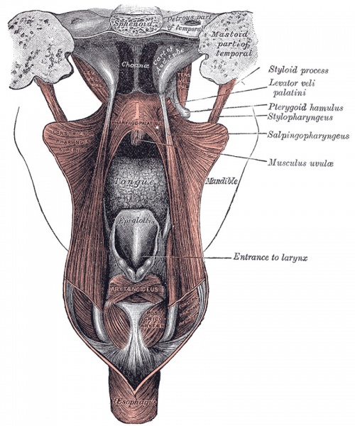
Fig. 1028 Dissection of the Muscles of the Palate from behind
Muscles of the Palate: Levator veli palatini, Tensor veli palatini, Musculus uvulæ, Glossopalatinus, Pharyngopalatinus.
Levator veli palatini
(Levator palati) is a thick, rounded muscle situated lateral to the choanæ. It arises from the under surface of the apex of the petrous part of the temporal bone and from the medial lamina of the cartilage of the auditory tube. After passing above the upper concave margin of the Constrictor pharyngis superior it spreads out in the palatine velum, its fibers extending obliquely downward and medialward to the middle line, where they blend with those of the opposite side.
Tensor veli palatini
(Tensor palati) is a broad, thin, ribbon-like muscle placed lateral to the Levator veli palatini. It arises by a flat lamella from the scaphoid fossa at the base of the medial pterygoid plate, from the spina angularis of the sphenoid and from the lateral wall of the cartilage of the auditory tube. Descending vertically between the medial pterygoid plate and the Pterygoideus internus it ends in a tendon which winds around the pterygoid hamulus, being retained in this situation by some of the fibers of origin of the Pterygoideus internus. Between the tendon and the hamulus is a small bursa. The tendon then passes medialward and is inserted into the palatine aponeurosis and into the surface behind the transverse ridge on the horizontal part of the palatine bone.
Musculus uvulæ
(Azygos uvulæ) arises from the posterior nasal spine of the palatine bones and from the palatine aponeurosis; it descends to be inserted into the uvula.
Glossopalatinus
(Palatoglossus) is a small fleshy fasciculus, narrower in the middle than at either end, forming, with the mucous membrane covering its surface, the glossopalatine arch. It arises from the anterior surface of the soft palate, where it is continuous with the muscle of the opposite side, and passing downward, forward, and lateralward in front of the palatine tonsil, is inserted into the side of the tongue, some of its fibers spreading over the dorsum, and others passing deeply into the substance of the organ to intermingle with the Transversus linguæ.
Pharyngopalatinus
(Palatopharyngeus) is a long, fleshy fasciculus narrower in the middle than at either end, forming, with the mucous membrane covering its surface, the pharyngopalatine arch. It is separated from the Glossopalatinus by an angular interval, in which the palatine tonsil is lodged. It arises from the soft palate, where it is divided into two fasciculi by the Levator veli palatini and Musculus uvulæ. The posterior fasciculus lies in contact with the mucous membrane, and joins with that of the opposite muscle in the middle line; the anterior fasciculus, the thicker, lies in the soft palate between the Levator and Tensor, and joins in the middle line the corresponding part of the opposite muscle. Passing lateralward and downward behind the palatine tonsil, the Pharyngopalatinus joins the Stylopharyngeus, and is inserted with that muscle into the posterior border of the thyroid cartilage, some of its fibers being lost on the side of the pharynx and others passing across the middle line posteriorly, to decussate with the muscle of the opposite side.
- Links: Palate Development
- Gray's Images: Development | Lymphatic | Neural | Vision | Hearing | Somatosensory | Integumentary | Respiratory | Gastrointestinal | Urogenital | Endocrine | Surface Anatomy | iBook | Historic Disclaimer
| Historic Disclaimer - information about historic embryology pages |
|---|
| Pages where the terms "Historic" (textbooks, papers, people, recommendations) appear on this site, and sections within pages where this disclaimer appears, indicate that the content and scientific understanding are specific to the time of publication. This means that while some scientific descriptions are still accurate, the terminology and interpretation of the developmental mechanisms reflect the understanding at the time of original publication and those of the preceding periods, these terms, interpretations and recommendations may not reflect our current scientific understanding. (More? Embryology History | Historic Embryology Papers) |
| iBook - Gray's Embryology | |
|---|---|

|
|
Reference
Gray H. Anatomy of the human body. (1918) Philadelphia: Lea & Febiger.
Cite this page: Hill, M.A. (2024, April 19) Embryology Gray1028.jpg. Retrieved from https://embryology.med.unsw.edu.au/embryology/index.php/File:Gray1028.jpg
- © Dr Mark Hill 2024, UNSW Embryology ISBN: 978 0 7334 2609 4 - UNSW CRICOS Provider Code No. 00098G
File history
Click on a date/time to view the file as it appeared at that time.
| Date/Time | Thumbnail | Dimensions | User | Comment | |
|---|---|---|---|---|---|
| current | 17:16, 28 April 2011 |  | 800 × 960 (202 KB) | S8600021 (talk | contribs) |
You cannot overwrite this file.
File usage
The following 2 pages use this file:
